PERIPHERAL VASCULAR DISEASE/PERIPHERAL ARTERY DISEASE (PAD)
Intermittent claudication = pain in the limb brought on by exertion and relieved at rest. This will recur on similar
effort (reproducible). The muscle isn’t getting as much oxygen as it requires so it causes pain/cramping.
Critical limb ischaemia = pain in the limb at rest (constant) with or without tissue loss. This will also produce a
BP reading of <50 mmHg at the ankle. These patients will often require opiate pain relief. The blood flow and
oxygen delivery is so little it causes pain at rest, often at night.
PATHOPHYSIOLOGY
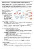
The main process leading up to peripheral
vascular/artery disease (PAD) is
atherosclerosis.
Arteriosclerosis → Atherosclerosis
= thickening of the arterial wall,
which causes loss of elasticity
▪ Focal disease of the large and
medium sized arteries
▪ Clinical manifestations include:
CAD, cerebrovascular disease,
peripheral artery disease
▪ Due to formation of fatty plaques in
arterial walls.
There are 3 stages of atherosclerotic development.
1. Endothelial damage
▪ The endothelium of an artery is involve in maintaining vasomotor tone, and does so by vasodilators (NO,
PGI2) and vasoconstrictors (angiotensin II, endothelin).
▪ It is also involved in thrombosis and leukocyte interaction, and it does so by expressing cellular adhesion
molecules.
▪ Therefore, if this endothelium is damaged, these functions are impaired and it is thus dysfunctional – it
will display inappropriate vasoconstriction and anti-thrombotic properties.
▪ Damage can be caused by: shear stress, toxicity, hyperlipidaemia, infection.
2. Formation of foam cells (infiltration of macrophages)
▪ Once the endothelium is damaged, oxidised LDLs can pass into it.
o Oxidation is facilitated by ROS from cellular damage
o Glycation can also occur, this is facilitated by high glucose levels. Glycated LDLs are more likely
to be oxidised.
▪ Diabetics will therefore have higher glycated LDL numbers.
▪ These are taken up by the scavenger receptor.
o Usually, unmodified LDLs are taken up by the LDL receptor, which recognises apolipoprotein
B100. Negative feedback occurs and once the LDLs are internalised, then surface LDL receptors
decrease in order to decrease LDL uptake.
o But modified LDLs aren’t recognised by the LDL receptor.
o There is no negative feedback of the scavenger receptor, so uptake of modified LDLs is
unlimited.
▪ The oxidised LDLs in the endothelial wall stimulate expression of inflammatory mediators (including
monocyte adhesion molecules).
▪ This attracts monocytes, which can bind to and cross the endothelium, where they become
macrophages.
▪ Macrophages can then accumulate the oxidised LDLs to become foam cells (these appear as fatty
streaks).
3. Fibrous cap formation
▪ Endothelial cells and macrophages release growth factors (platelet derived growth factor/PDGF).
▪ PGDF causes proliferation of smooth muscle cells in the tunica intima of the artery, as well as collagen
production.
▪ This proliferation causes atrophy of the internal elastic lamina, and the artery becomes loses elasticity.
▪ These smooth muscle cells can also become foam cells as they can also take up modified LDLs.
Intermittent claudication = pain in the limb brought on by exertion and relieved at rest. This will recur on similar
effort (reproducible). The muscle isn’t getting as much oxygen as it requires so it causes pain/cramping.
Critical limb ischaemia = pain in the limb at rest (constant) with or without tissue loss. This will also produce a
BP reading of <50 mmHg at the ankle. These patients will often require opiate pain relief. The blood flow and
oxygen delivery is so little it causes pain at rest, often at night.
PATHOPHYSIOLOGY
The main process leading up to peripheral
vascular/artery disease (PAD) is
atherosclerosis.
Arteriosclerosis → Atherosclerosis
= thickening of the arterial wall,
which causes loss of elasticity
▪ Focal disease of the large and
medium sized arteries
▪ Clinical manifestations include:
CAD, cerebrovascular disease,
peripheral artery disease
▪ Due to formation of fatty plaques in
arterial walls.
There are 3 stages of atherosclerotic development.
1. Endothelial damage
▪ The endothelium of an artery is involve in maintaining vasomotor tone, and does so by vasodilators (NO,
PGI2) and vasoconstrictors (angiotensin II, endothelin).
▪ It is also involved in thrombosis and leukocyte interaction, and it does so by expressing cellular adhesion
molecules.
▪ Therefore, if this endothelium is damaged, these functions are impaired and it is thus dysfunctional – it
will display inappropriate vasoconstriction and anti-thrombotic properties.
▪ Damage can be caused by: shear stress, toxicity, hyperlipidaemia, infection.
2. Formation of foam cells (infiltration of macrophages)
▪ Once the endothelium is damaged, oxidised LDLs can pass into it.
o Oxidation is facilitated by ROS from cellular damage
o Glycation can also occur, this is facilitated by high glucose levels. Glycated LDLs are more likely
to be oxidised.
▪ Diabetics will therefore have higher glycated LDL numbers.
▪ These are taken up by the scavenger receptor.
o Usually, unmodified LDLs are taken up by the LDL receptor, which recognises apolipoprotein
B100. Negative feedback occurs and once the LDLs are internalised, then surface LDL receptors
decrease in order to decrease LDL uptake.
o But modified LDLs aren’t recognised by the LDL receptor.
o There is no negative feedback of the scavenger receptor, so uptake of modified LDLs is
unlimited.
▪ The oxidised LDLs in the endothelial wall stimulate expression of inflammatory mediators (including
monocyte adhesion molecules).
▪ This attracts monocytes, which can bind to and cross the endothelium, where they become
macrophages.
▪ Macrophages can then accumulate the oxidised LDLs to become foam cells (these appear as fatty
streaks).
3. Fibrous cap formation
▪ Endothelial cells and macrophages release growth factors (platelet derived growth factor/PDGF).
▪ PGDF causes proliferation of smooth muscle cells in the tunica intima of the artery, as well as collagen
production.
▪ This proliferation causes atrophy of the internal elastic lamina, and the artery becomes loses elasticity.
▪ These smooth muscle cells can also become foam cells as they can also take up modified LDLs.