Written by students who passed Immediately available after payment Read online or as PDF Wrong document? Swap it for free 4.6 TrustPilot
logo-home
Document preview thumbnail
Preview 2 out of 8 pages
Essay

Anatomy: Respiratory Assignment Exemplar

Document preview thumbnail
Preview 2 out of 8 pages

Unit 1: Assignment 4 Components of the Respiratory System, structure and function of the lungs, mechanics of breathing, respiratory volumes, control of breathing and gaseous exchange. Submitted for my BTEC assignment and achieved a Distinction.

Content preview

Unit 1 – Anatomy

Assignment 4 – Respiratory System

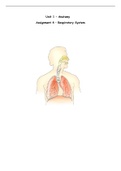
, The Respiratory System diagram

Epiglottis/
Nasal
Pharynx
cavity

Larynx

Trachea

Lungs


Bronchioles
Bronchus
Alveoli


Diaphragm
Nasal cavity – This starts in the nose, and leads to the epiglottis. It is lined with hair to filter
the air, removing debris, and to moisten the air.

Epiglottis/Pharynx - The epiglottis is a flexible flap at the start of the larynx. It acts as a
switch between the larynx and the oesophagus. It protects you from choking on food that could
obstruct the airway.

Larynx - The larynx is a tough, flexible part of the respiratory tract connecting the pharynx to
the trachea in the neck. It plays a vital role in the respiratory tract by allowing air to pass
through it while keeping food and drink from blocking the airway. The larynx is also the body’s
“voice box” as it contains the vocal folds that produce the sounds of speech and singing.

Trachea - The trachea, also called the windpipe, filters the air that is inhaled. It branches into
the bronchi, which are two tubes that carry air into each lung.

Bronchus – This is where the air enters the lungs. It is the biggest passageway, and it send air
into the left and right lung. They branch off into the bronchioles, smaller tubes.

Bronchioles – These are small tubes that split off from the bronchus. There are lots of them to
make the respiration process more efficient. It means that more air will get sent to the alveoli.

Alveoli – These are located at the end of the bronchioles. They are tiny air sacs that allow the
exchange of gases. Surrounding the alveoli, there is a dense network of capillaries to allow to
exchange. Capillaries are one cell thick, and the alveoli are thin walled to allow the diffusion of
gases. There are loads of bronchioles and alveoli so that the air can be spread out, to lower
blood pressure, so they don’t burst or break.

Diaphragm – This is located underneath the lungs. It is a respiratory muscle, the main muscle
involved in breathing at rest. It is dome-shaped, which determines the space in the thoracic
cavity. It contracts and moves down during inspiration, and relaxes and moves up during
expiration.

Document information

Study Level
Subject
Uploaded on
April 11, 2023
Number of pages
8
Written in
2015/2016
Type
Essay
Professor(s)
Unknown
Grade
A+
$8.33

Wrong document? Swap it for free Within 14 days of purchase and before downloading, you can choose a different document. You can simply spend the amount again.
Written by students who passed
Immediately available after payment
Read online or as PDF

Sold
1
Followers
1
Items
12
Last sold
3 year ago


Why students choose Stuvia

Created by fellow students, verified by reviews

Quality you can trust: written by students who passed their tests and reviewed by others who've used these notes.

Didn't get what you expected? Choose another document

No worries! You can instantly pick a different document that better fits what you're looking for.

Pay as you like, start learning right away

No subscription, no commitments. Pay the way you're used to via credit card and download your PDF document instantly.

Student with book image

“Bought, downloaded, and aced it. It really can be that simple.”

Alisha Student

Working on your references?

Create accurate citations in APA, MLA and Harvard with our free citation generator.

Working on your references?

Frequently asked questions