

4. ONDERSTE LIDMAAT
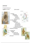
4.1 Heupgewricht (articulatio coxae)
(=kogelgewricht) → pwp 1 vanaf dia 13 caput femoris en acetabulum
4.1.1 Gewrichtskapsel
- membrana synovialis
- omringt gewrichtsholte:
- proximaal labrum
- distaal: collum femoris
- concave wand vd fac lunata → (bij
incisura acetabuli) → lig capitis femoris
- membrana fibrosa
- gedraaid vezelverloop
- proximaal: rondom labrum
- distaal: voorbij collum femoris
- ventraal: linea intertrochanterica
- dorsaal: mediaal van crista intertrochanterica
- fossa trochanterica en beide fossa’s vallen
buiten kapsel (daar zijn er veel spierinserties)
4.1.2 Versterkingen vh kapsel
- zona orbicularis
- band rond smalste deel v collum
- bedekt door andere ligamenten
- collum kan draaien, caput tegen
gewrichtspan geduwd
1
, - kan er =/= uit zonder zona te scheuren
- eigen vezels + eindvezels lig. pubofemorale en ischiofemorale
- ligamentum iliofemorale(ligament van Bertin)
- = stevigste
- ventraal: kapsel (rand vh acetabulum)
- spina illiaca anterior + gladde been lateraal daarvan + acetabulum
- loop: driehoekig over voorzijde van capsula articularis
- I: linea intertrochanterica
- pars lateralis/ superior= bovenrand collum naar linea intertrochanterica thv trochanter major
- pars medialis/ inferior= ventrale zijde vh collum naar linea intertrochanterica thv trochanter minor
- functie: anteflexie, remt retroflexie
- gesloten: ontspanning bij retroflexie
- remt dorsaal kantelen tov OL
- rechtopstaan → lig iliofemorale (=/= dorsaal kantelen) + tonus vd spieren vd regio glutea (=/= ventraal kantelen)
- remt exorotatie (en remt ook licht endorotatie)
- pars superior: ontspannen bij abductie en gespannen bij adductie
- ligamentum pubofemorale
- ventraal: kapsel (rand vh acetabulum + eminentia iliopubica)
- V: voorzijde capusla, lateraal over collum femoris
- I: linea intertrochanterica boven trochanter minor (bij pars inf vh lig iliofem)
- veel vezels gaan over in zona orbicularis
- functie: remt abductie
- ligamentum ischiofemorale
- O: dorsaal op os ischii + dorsaal acetabulum
- V: achter caput, over collum
- I: mediaal trochanter major (bij pars sup vh lig iliofem)
- veel vezels gaan over in zona orbicularis
- functie: versterkt achterzijde vh gewricht
- remt retroflexie (belet achteroverkantelen bij staan)
- remt endorotatie
- anteflexie en endorotatie
2
, 4.1.3 Bewegingen vd dij in heupgewricht
- (pelvis = gefixeerd, dij = beweeglijk) → bewegingen van bekken uitsluiten
- anteflexie(120°)+ retroflexie(13°)rond de laterolateraleas
- retroflexie : geremd door alle banden (vooral onderste deel vh lig iliofem + lig ischiofem)
- in rechtopstaande houding kan het bekken nauwelijks achterover gekanteld worden
- anteflexie : geremd door dorsale dijspieren (vooral bij gestrekte knie)
- bij gebogen knie kan dij tot buikwand
- abductie(40°)+ adductie(20°) rond de anteroposterieureas
- endorotatie(36°)+ exorotatie(13°)rond de longitudinaleas
- circumductie (= combinatie van de 3)
- stabilisatie:
- pars sup vh lig iliofem remt endo, exorotatie en adductie
- pars inf vh lig iliofem remt exorotatie
- lig pubofem remt abductie en exorotatie
- lig ischiofem remt endorotatie
4.1.4 Bewegingen vh bekken ih heupgewricht
- (dij = gefixeerd, pelvis = beweeglijk) → bewegingen vh dijbeen uitsluiten
- referentiepunt = spina iliaca anterior superior
- anteversie= voorwaarts kantelen → toename lendenlordose
- retroversie= achterwaarts kantelen → afname lendenlordose
- lateroflexie = ene kant vh bekken daalt (andere zijdenaar craniaal) → lateroflexie vd wervelkolom
- rotatie mediaal/lateraal→ zijn maar beperkt mogelijk
4.2 Kniegewricht (articulatio genus)
( = (draai)scharniergewricht) → pwp 1 vanaf dia 17
tibia: facies articularis superiores + eminentia intercondylaris
femur: condylen + facies patellaris
3
, patella: facies articularis
→ alle gewrichtsvlakken zijn bekleed met hyalien kraakbeen
4.2.1 Menisci
- tss gewrichtsopp vd tibia en de femur: menisci articulares
- structuur: sterk bindweefsel, opp = vezelkraakbeen, diepte = peesweefsel
- periferie = breed (diepe zijde capsula articularis) → dunne boord in midden
- centrum = volledig open
- meniscus lateralis = ⅘ gesloten O
- meniscus medialis = omgekeerde C
- OECI: O extern; C intern
- ligging: ventraal en dorsaal vd eminentia intercondylaris op tibia
- hebben beide nog een aanhechting
- mediaal: ligamentum collaterale tibiale
- lateraal: oorsprongspees vd M.popliteus
- tss men lateralis en femur → (achteraan in capsula articularis) → ligamentum meniscofemorale
posterius (=afspliting vd achterste kruisband)
- vervorming bij beweging
- mediaal: bij rotatie (dus tss menisci en tibia)
- lateraal: bij bij bewegingen vd laterale conylus, M. popliteus-pees, lig meniscofemorale posterius
- verbonden door ligamentum transversum genu
- onvolledige horizontale scheidingswand tss craniaal en caudaal in gewricht
- craniaal = concaaf → gewrichtspan (= articulatio meniscofemoralis) → articulatie met condylen van femur
- caudaal = vlak → (articulatio meniscotibialis) → articulatie met licht concave facies articulares superiores
vd tibia
4.2.2 Ligamenta cruciata genu (=kruisbanden)
- onvolledig verticaal tussenschot tss het laterale en mediale gedeelte
- ligamentum cruciatum anterius
4
4.1 Heupgewricht (articulatio coxae)
(=kogelgewricht) → pwp 1 vanaf dia 13 caput femoris en acetabulum
4.1.1 Gewrichtskapsel
- membrana synovialis
- omringt gewrichtsholte:
- proximaal labrum
- distaal: collum femoris
- concave wand vd fac lunata → (bij
incisura acetabuli) → lig capitis femoris
- membrana fibrosa
- gedraaid vezelverloop
- proximaal: rondom labrum
- distaal: voorbij collum femoris
- ventraal: linea intertrochanterica
- dorsaal: mediaal van crista intertrochanterica
- fossa trochanterica en beide fossa’s vallen
buiten kapsel (daar zijn er veel spierinserties)
4.1.2 Versterkingen vh kapsel
- zona orbicularis
- band rond smalste deel v collum
- bedekt door andere ligamenten
- collum kan draaien, caput tegen
gewrichtspan geduwd
1
, - kan er =/= uit zonder zona te scheuren
- eigen vezels + eindvezels lig. pubofemorale en ischiofemorale
- ligamentum iliofemorale(ligament van Bertin)
- = stevigste
- ventraal: kapsel (rand vh acetabulum)
- spina illiaca anterior + gladde been lateraal daarvan + acetabulum
- loop: driehoekig over voorzijde van capsula articularis
- I: linea intertrochanterica
- pars lateralis/ superior= bovenrand collum naar linea intertrochanterica thv trochanter major
- pars medialis/ inferior= ventrale zijde vh collum naar linea intertrochanterica thv trochanter minor
- functie: anteflexie, remt retroflexie
- gesloten: ontspanning bij retroflexie
- remt dorsaal kantelen tov OL
- rechtopstaan → lig iliofemorale (=/= dorsaal kantelen) + tonus vd spieren vd regio glutea (=/= ventraal kantelen)
- remt exorotatie (en remt ook licht endorotatie)
- pars superior: ontspannen bij abductie en gespannen bij adductie
- ligamentum pubofemorale
- ventraal: kapsel (rand vh acetabulum + eminentia iliopubica)
- V: voorzijde capusla, lateraal over collum femoris
- I: linea intertrochanterica boven trochanter minor (bij pars inf vh lig iliofem)
- veel vezels gaan over in zona orbicularis
- functie: remt abductie
- ligamentum ischiofemorale
- O: dorsaal op os ischii + dorsaal acetabulum
- V: achter caput, over collum
- I: mediaal trochanter major (bij pars sup vh lig iliofem)
- veel vezels gaan over in zona orbicularis
- functie: versterkt achterzijde vh gewricht
- remt retroflexie (belet achteroverkantelen bij staan)
- remt endorotatie
- anteflexie en endorotatie
2
, 4.1.3 Bewegingen vd dij in heupgewricht
- (pelvis = gefixeerd, dij = beweeglijk) → bewegingen van bekken uitsluiten
- anteflexie(120°)+ retroflexie(13°)rond de laterolateraleas
- retroflexie : geremd door alle banden (vooral onderste deel vh lig iliofem + lig ischiofem)
- in rechtopstaande houding kan het bekken nauwelijks achterover gekanteld worden
- anteflexie : geremd door dorsale dijspieren (vooral bij gestrekte knie)
- bij gebogen knie kan dij tot buikwand
- abductie(40°)+ adductie(20°) rond de anteroposterieureas
- endorotatie(36°)+ exorotatie(13°)rond de longitudinaleas
- circumductie (= combinatie van de 3)
- stabilisatie:
- pars sup vh lig iliofem remt endo, exorotatie en adductie
- pars inf vh lig iliofem remt exorotatie
- lig pubofem remt abductie en exorotatie
- lig ischiofem remt endorotatie
4.1.4 Bewegingen vh bekken ih heupgewricht
- (dij = gefixeerd, pelvis = beweeglijk) → bewegingen vh dijbeen uitsluiten
- referentiepunt = spina iliaca anterior superior
- anteversie= voorwaarts kantelen → toename lendenlordose
- retroversie= achterwaarts kantelen → afname lendenlordose
- lateroflexie = ene kant vh bekken daalt (andere zijdenaar craniaal) → lateroflexie vd wervelkolom
- rotatie mediaal/lateraal→ zijn maar beperkt mogelijk
4.2 Kniegewricht (articulatio genus)
( = (draai)scharniergewricht) → pwp 1 vanaf dia 17
tibia: facies articularis superiores + eminentia intercondylaris
femur: condylen + facies patellaris
3
, patella: facies articularis
→ alle gewrichtsvlakken zijn bekleed met hyalien kraakbeen
4.2.1 Menisci
- tss gewrichtsopp vd tibia en de femur: menisci articulares
- structuur: sterk bindweefsel, opp = vezelkraakbeen, diepte = peesweefsel
- periferie = breed (diepe zijde capsula articularis) → dunne boord in midden
- centrum = volledig open
- meniscus lateralis = ⅘ gesloten O
- meniscus medialis = omgekeerde C
- OECI: O extern; C intern
- ligging: ventraal en dorsaal vd eminentia intercondylaris op tibia
- hebben beide nog een aanhechting
- mediaal: ligamentum collaterale tibiale
- lateraal: oorsprongspees vd M.popliteus
- tss men lateralis en femur → (achteraan in capsula articularis) → ligamentum meniscofemorale
posterius (=afspliting vd achterste kruisband)
- vervorming bij beweging
- mediaal: bij rotatie (dus tss menisci en tibia)
- lateraal: bij bij bewegingen vd laterale conylus, M. popliteus-pees, lig meniscofemorale posterius
- verbonden door ligamentum transversum genu
- onvolledige horizontale scheidingswand tss craniaal en caudaal in gewricht
- craniaal = concaaf → gewrichtspan (= articulatio meniscofemoralis) → articulatie met condylen van femur
- caudaal = vlak → (articulatio meniscotibialis) → articulatie met licht concave facies articulares superiores
vd tibia
4.2.2 Ligamenta cruciata genu (=kruisbanden)
- onvolledig verticaal tussenschot tss het laterale en mediale gedeelte
- ligamentum cruciatum anterius
4


