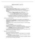
NURS 300 wk 1-2
Med Surg wks. 1-2. Q1
Important Lab Ranges / Values
Electrolyte Labs Normal
Potassium 3.5 - 5 mEq/L
Sodium 136 to 145 mEq/L
Magnesium 1.3 to 2.1 mEq/L
Calcium 9 to 10.5 mg/dL
Phosphorus 3 - 4.5 mg/dL
Chloride 98 – 106 mEq/L
Glucose: 60-120 mg/dL Therapeutic PT:1.5-2 x control = 14.25-24 seconds
RBC Male:4.6-6.2 million/uL Therapeutic PTT: 24-100 seconds
RBC Female: 4.2-5.4 million/uL Albumin: 3.5-5.0 g/dL
WBC: 5,000-10,000 uL Bilirubin: 0.3-1.0 mg/dL (Liver function).
HGB Male: 14-18 g/dL Ammonia: 0.3-1.0 mg/dL
HGB Female: 12-16 g/dL
Protein: 3-5 g/dL
HCT Male:42-52%
Urine pH: 4.6-8
HCT Female: 37-47%
Creatine in Males: 0.6-1.2 mg/dL
PTT: 30-45 seconds (monitor heparin therapy).
Platelets: 150,000-400,000/mm3 Creatine in females: 0.5-1.1 mg/dL (Kidney function).
PT: 11-12.5 seconds (monitor Warfarin Therapy). BUN: 10-20 mg/dL (Renal function).
INR: 2.0-3.0 Therapeutic range Digoxin level: 0.8-2 mg/dL
Lithium: 0.4-1.0 mEq/L
Health and Wellness 1. Modifiable: Can be changed.
a. Ex: Smoking, diet, lifestyle
2. Nonmodifiable: Cannot be changed
a. Age, sex, genetics
Illness This is an altered level of functioning in response to a disease process.
1. Response to illness can be influenced by:
a. Degree of physical changes
b. Perceptions by self
c. Cultural values & beliefs
d. Denial or fears of illness
e. Social demands, time constraints, economic resources, and health care access.
Disease This is a condition that results in the physiological alteration in the composition of the body
Nursing care Evaluate the health needs of a client and create strategies to meet those needs.
Provide resources
Identify and encourage the use of support systems
Interventions: Identify obstacles to health and wellness and create strategies to reduce these obstacles
Identify ways to reduce health risks
Develop health education methods to improve health awareness and reduce health
risks.
Cardiovascular
, The cardiovascular (CV) system is responsible for supplying oxygen to body organs and other tissues
(perfusion). It is made up of the heart and blood vessels (both arteries and veins).
Cardiac Function:
Cardiac
Function Systole: The Ventricles and Atria contract and eject the blood.
Diastole: During Diastole, this is where the ventricles and the atria are
relaxed and filling up with blood.
Within the cardiac cycle: 2/3 of the time is spent in diastole and 1/3 of the
time is spent in systole.
The percentage of blood leaving the left ventricle each time it contracts is
called?
a. Left ventricular ejection fraction
Extra notes:
S1 is the first sound, which occurs when the mitral and tricuspid valves close at the
beginning of systole (when the heart contracts).
S2 is the second sound, which occurs when the aortic and pulmonary valves close at the
end of systole (when the heart finishes contracting).
Volume of blood in the ventricles at the end of diastole (Right before
Preload contraction).
----This determines the amount of stretch that we are placing on those
myocardial fibers in the heart.
Is the Peripheral resistance that the left ventricle must overcome in order
to push the blood into systemic circulation.
When caring for patients with heart failure, we will be administering
Afterload medications often that will reduce the preload and or afterload, to try to
reduce the amount of stretch in the heart in the amount or workload the
heart needs to do by reducing preload, and also trying to reduce afterload
to make it easier on the heart to push the blood into the systemic
circulation
Apical Pulse The pulse located at the left fifth intercostal space in the midclavicular line (in the
mitral area); also called the point of maximal impulse (PMI).
Sensory receptors in the arch of the aorta and at the origin of the internal carotid
Baroreceptors arteries that are stimulated when the arterial walls are stretched by an increased
blood pressure.
Mean arterial The arterial blood pressure necessary (between 60 and 70 mm Hg) necessary to
pressure maintain perfusion of major body organs, such as the kidneys and the brain.
MAP
Stroke Volume The amount of blood ejected by the left ventricle during each contraction.
The amount of pressure or force generated by the left ventricle to distribute blood into the aorta
Systolic BP with each contraction of the heart. Ventricular contraction.
Systole: The phase of the cardiac cycle that consists of the contraction and emptying of the atria and
ventricles.
, The amount of pressure or force against the arterial walls during the relaxation
Diastolic BP phase of the cardiac cycle.
Diastole: The phase of the cardiac cycle that consists of relaxation and filling of
the atria and ventricles; normally about two-thirds of the cardiac cycle.
Troponin A myocardial muscle protein released into the bloodstream with injury to
myocardial muscle.
Cardiac output:
How to calculate Is the volume of blood in liters that is ejected from the left ventricle every
minute.
cardiac output Cardiac Output = CO = HR x SV
HR = is the number of times that the ventricles contract within a minute
Heart rate x (60-100 bpm).
Stroke Volume Stroke Volume = Volume of blood in liters that is ejected from the ventricle
with each heartbeat (4-8 liters per minute).
Systole = Contraction
Diastole = Relaxation
Pulse Pressure The difference between systolic and diastolic BP
Hypotension When a client experiences hypotension, baroreceptors in the aortic arch sense a pressure
decrease in the vessels. The parasympathetic system responds by lessening the inhibitory
effect on the sinoatrial node. This results in an increase in heart rate and respiratory rate.
This tachycardia is an early response and is seen even when blood pressure is not critically
low. An increased heart rate and respiratory rate will compensate for the low blood
pressure and maintain oxygen saturation and perfusion. The client may not be able to
compensate for long and decreased oxygenation and cool, clammy skin will occur later.
The P wave represents the depolarization of the atria.
Waves of the The P-R interval represents depolarization of the atria, atrioventricular (AV) node,
heart bundle of His, bundle branches, and the Purkinje fibers.
The QRS represents ventricular depolarization. Normal QRS interval is <0.12
The Q-T interval represents depolarization and repolarization of the entire
conduction system.
P wave, of the rhythm strip to evaluate for atrial depolarization.
the QRS complex, of the rhythm strip to evaluate for ventricular depolarization.
the T wave, of the rhythm strip to evaluate for ventricular repolarization.
Bradydysrhythmia An abnormal heart rhythm with a heart rate less than 60 beats/min; also known
as bradyarrhythmia.
Bradydysrhythmia can cause decreased systemic perfusion, which can lead to
confusion. Therefore, the nurse should monitor the client's mental status.
Pericarditis inflammation or alteration of the pericardium (the membranous sac that encloses
the heart).
A client who has pericarditis will experience dyspnea, hiccups and nonproductive
cough. These manifestations can indicate HF from pericardial compression due to
constrictive pericarditis or cardiac tamponade