

BIOL 235 FINAL EXAM NOTES
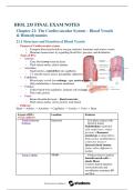
Chapter 21: The Cardiovascular System – Blood Vessels
& Hemodynamics
21.1 Structure and Function of Blood Vessels
• Purpose of Cardiovascular system:
• Transport blood and to deliver oxygen, nutrients, hormones and remove wastes
• Maintains homeostasis by regulating blood flow, pressure, and distribution
• Types of BVs:
• Arteries:
• Carry blood away from the heart
• Thick tunica media, elastic laminae
• Arterioles:
• Small arteries, control flow into capillaries
• 1-2 smooth muscle layers, precapillary sphincters
• Capillaries:
• Microscopic vessels for exchange – gas, nutrient, waste
• Only endothelium + basement membrane
• Venules:
• Collect blood from capillaries, immune cell exchange
• Thin walls, porous
• Veins:
• Return blood to the heart – blood reservoirs
• Thick tunica media, valves present, wide lumen
• Pathway:
Heart -> Arteries -> Arterioles -> Capillaries -> Venules -> Veins -> Heart
Layers of BVs Location Features
(except capillaries)
• Tunica Interna Innermost • Is in direct contact with
(Intima) blood in lumen
Endothelium (epithelial
cells single layer; reduce
friction) + Basement
membrane (collage fibers;
helps anchor endothelium to
connective tissue) +
Internal elastic lamina
(holes; allow diffusion to
media)
• Tunica Media Middle • Thickest layer in arteries
Smooth muscle cells +
elastic fibers
Controls lumen diameter:
vasoconstriction and
vasodilation; maintains BP
and flow
, External elastic lamina
(separates media & externa)
• Tunica Externa Outermost Collagen & elastic fibers +
(Adventitia) nerve fibers; contains vasa
vasorum
• Vas vasorum:
Tiny BVs that supply the
walls of larger BVs
• Anchors BV to tissue
Arteries
• Stretch easily in response to small increase in pressure
• Carry blood away from the heart
• Thick tunica media
• Elasticity & contractility are key
• Elasticity allows compliance => absorbs pressure
• Contractility regulates flow
• Types:
• Elastic Arteries (“Conducting arteries”)
• Largest arteries
• Aorta, pulmonary trunk
• High in elastic fibers in tunica media
• Elastic lamellae
• Function:
• Pressure reservoir
• Store mechanical energy and maintain blood flow during diastole
• Expand/stretch during systole
• Recoil during diastole to propel blood forward
• Aorta, brachiocephalic, subclavian, carotid, iliac arteries
• Muscular Arteries (“Distributing arteries”)
• Medium-sized
• Brachial, radial, femoral
• Thick smooth muscle layer -> vasoconstriction/dilation
• Less elastic than elastic arteries
• Have vascular tone: slight continuous contraction to stiffen vessel and maintain
pressure
• Function:
• Control blood flow to specific organs
• tunica externa thicker than tunica media
,Arterial Anastomoses
• Anastomoses:
• Union of two or more arteries supplying the same region
• Function:
• Provide collateral circulation (backup routes)
• Maintains perfusion during blockages or compressions
• Arteries that do not anastomoses = end arteries
• Blockage of an end artery = tissue necrosis (death)
Arterioles
• Smallest arteries (15-300 μm)
• Control flow into capillaries (resistance vessels)
• Structure:
• 1-2 smooth muscle layers
• Internal elastic lamina (disappears distally)
• Last muscle cell = precapillary sphincter
• High impact on systemic vascular resistance (SVR)
Capillaries (exchange vessels)
• Smallest vessels (5-10 μm)
• Wall: only endothelium + basement membrane
• Form capillary beds (~10-100 per arteriole)
• Types:
• Continuous:
• No pores
• Tight junctions
• CNS, skin muscle
• Fenestrated:
• Pores
• Kidneys, endocrine glands
• Sinusoids:
• Wide, with gaps
• Liver, red bone marrow
Venules
• Collect blood from capillaries
• Postcapillary venules (10-15 μm):
• Very porous
• Site of WBC emigration and exchange
• Muscular venules (50-200 μm):
• Smooth muscle present
• Start to resemble veins
Veins
• Return blood to the heart
• Wall: 3 tunics, but thin media, large lumen
- Valves:
• Thin folds of tunica interna that prevent backflow (important in limbs)
• Function:
• Blood reservoirs
, • ~64% of blood volume at rest is in systemic veins/venules
• Less elastic = cannot withstand high pressure
Summary
Vessel Type Size Internal Structure Function
Conduct blood; pressure
• Elastic Arteries Largest Elastic lamellae; thin wall
reservoir
• Muscular Smooth muscle; defined
Arteries Medium Distribute blood; control flow
laminae
Few smooth muscle layers; Control resistance, flow to
• Arterioles 15–300 μm sphincter
capillaries
• Capillaries 5–10 μm Endothelium only Exchange vessels
Collect blood; site of WBC
• Venules 10–200 μm Thin wall, few muscle cells
action
0.5 mm–3
• Veins cm Valves; large lumen Return blood; blood reservoir
21.2 Capillary Exchange
• The capillaries are the primary exchange sites for gases, nutrients, hormones, and
wastes between blood and body tissues
• At any given time, only ~7% of total blood is in systemic capillaries
• But this small amount is critical because of constant capillary exchange
Three Main Mechanisms of Capillary Exchange
1. Diffusion:
• (Most important)
• Passive movement down a concentration gradient (high -> low)
• Substances moved:
• O2, glucose, amino acids, and hormones (blood -> interstitial fluid)
• CO2, wastes (interstitial fluid -> blood)
• Routes of movement:
Through endothelial cell membranes
• E.g., lipid-soluble substances like O2, CO2, steroids
Through intercellular clefts or fenestrations
• E.g., water-soluble substances like glucose, ions
• Blood capillaries (Blood-Brain Barrier):
• Continuous capillaries sealed by tight junctions
• Very limited diffusion – protects CNS from harmful substances
• Exceptions: hypothalamus, pineal gland, pituitary (no barrier)
2. Transcytosis (selective, vesicle-mediated transport):
• Process:
1. Endocytosis on one side of endothelial cell
2. Transport in vesicle
3. Exocytosis on opposite side
• Used for large, lipid-insoluble molecules that cannot pass via diffusion
• Insulin crossing capillary wall
• Maternal antibodies entering fetal circulation through placenta
Chapter 21: The Cardiovascular System – Blood Vessels
& Hemodynamics
21.1 Structure and Function of Blood Vessels
• Purpose of Cardiovascular system:
• Transport blood and to deliver oxygen, nutrients, hormones and remove wastes
• Maintains homeostasis by regulating blood flow, pressure, and distribution
• Types of BVs:
• Arteries:
• Carry blood away from the heart
• Thick tunica media, elastic laminae
• Arterioles:
• Small arteries, control flow into capillaries
• 1-2 smooth muscle layers, precapillary sphincters
• Capillaries:
• Microscopic vessels for exchange – gas, nutrient, waste
• Only endothelium + basement membrane
• Venules:
• Collect blood from capillaries, immune cell exchange
• Thin walls, porous
• Veins:
• Return blood to the heart – blood reservoirs
• Thick tunica media, valves present, wide lumen
• Pathway:
Heart -> Arteries -> Arterioles -> Capillaries -> Venules -> Veins -> Heart
Layers of BVs Location Features
(except capillaries)
• Tunica Interna Innermost • Is in direct contact with
(Intima) blood in lumen
Endothelium (epithelial
cells single layer; reduce
friction) + Basement
membrane (collage fibers;
helps anchor endothelium to
connective tissue) +
Internal elastic lamina
(holes; allow diffusion to
media)
• Tunica Media Middle • Thickest layer in arteries
Smooth muscle cells +
elastic fibers
Controls lumen diameter:
vasoconstriction and
vasodilation; maintains BP
and flow
, External elastic lamina
(separates media & externa)
• Tunica Externa Outermost Collagen & elastic fibers +
(Adventitia) nerve fibers; contains vasa
vasorum
• Vas vasorum:
Tiny BVs that supply the
walls of larger BVs
• Anchors BV to tissue
Arteries
• Stretch easily in response to small increase in pressure
• Carry blood away from the heart
• Thick tunica media
• Elasticity & contractility are key
• Elasticity allows compliance => absorbs pressure
• Contractility regulates flow
• Types:
• Elastic Arteries (“Conducting arteries”)
• Largest arteries
• Aorta, pulmonary trunk
• High in elastic fibers in tunica media
• Elastic lamellae
• Function:
• Pressure reservoir
• Store mechanical energy and maintain blood flow during diastole
• Expand/stretch during systole
• Recoil during diastole to propel blood forward
• Aorta, brachiocephalic, subclavian, carotid, iliac arteries
• Muscular Arteries (“Distributing arteries”)
• Medium-sized
• Brachial, radial, femoral
• Thick smooth muscle layer -> vasoconstriction/dilation
• Less elastic than elastic arteries
• Have vascular tone: slight continuous contraction to stiffen vessel and maintain
pressure
• Function:
• Control blood flow to specific organs
• tunica externa thicker than tunica media
,Arterial Anastomoses
• Anastomoses:
• Union of two or more arteries supplying the same region
• Function:
• Provide collateral circulation (backup routes)
• Maintains perfusion during blockages or compressions
• Arteries that do not anastomoses = end arteries
• Blockage of an end artery = tissue necrosis (death)
Arterioles
• Smallest arteries (15-300 μm)
• Control flow into capillaries (resistance vessels)
• Structure:
• 1-2 smooth muscle layers
• Internal elastic lamina (disappears distally)
• Last muscle cell = precapillary sphincter
• High impact on systemic vascular resistance (SVR)
Capillaries (exchange vessels)
• Smallest vessels (5-10 μm)
• Wall: only endothelium + basement membrane
• Form capillary beds (~10-100 per arteriole)
• Types:
• Continuous:
• No pores
• Tight junctions
• CNS, skin muscle
• Fenestrated:
• Pores
• Kidneys, endocrine glands
• Sinusoids:
• Wide, with gaps
• Liver, red bone marrow
Venules
• Collect blood from capillaries
• Postcapillary venules (10-15 μm):
• Very porous
• Site of WBC emigration and exchange
• Muscular venules (50-200 μm):
• Smooth muscle present
• Start to resemble veins
Veins
• Return blood to the heart
• Wall: 3 tunics, but thin media, large lumen
- Valves:
• Thin folds of tunica interna that prevent backflow (important in limbs)
• Function:
• Blood reservoirs
, • ~64% of blood volume at rest is in systemic veins/venules
• Less elastic = cannot withstand high pressure
Summary
Vessel Type Size Internal Structure Function
Conduct blood; pressure
• Elastic Arteries Largest Elastic lamellae; thin wall
reservoir
• Muscular Smooth muscle; defined
Arteries Medium Distribute blood; control flow
laminae
Few smooth muscle layers; Control resistance, flow to
• Arterioles 15–300 μm sphincter
capillaries
• Capillaries 5–10 μm Endothelium only Exchange vessels
Collect blood; site of WBC
• Venules 10–200 μm Thin wall, few muscle cells
action
0.5 mm–3
• Veins cm Valves; large lumen Return blood; blood reservoir
21.2 Capillary Exchange
• The capillaries are the primary exchange sites for gases, nutrients, hormones, and
wastes between blood and body tissues
• At any given time, only ~7% of total blood is in systemic capillaries
• But this small amount is critical because of constant capillary exchange
Three Main Mechanisms of Capillary Exchange
1. Diffusion:
• (Most important)
• Passive movement down a concentration gradient (high -> low)
• Substances moved:
• O2, glucose, amino acids, and hormones (blood -> interstitial fluid)
• CO2, wastes (interstitial fluid -> blood)
• Routes of movement:
Through endothelial cell membranes
• E.g., lipid-soluble substances like O2, CO2, steroids
Through intercellular clefts or fenestrations
• E.g., water-soluble substances like glucose, ions
• Blood capillaries (Blood-Brain Barrier):
• Continuous capillaries sealed by tight junctions
• Very limited diffusion – protects CNS from harmful substances
• Exceptions: hypothalamus, pineal gland, pituitary (no barrier)
2. Transcytosis (selective, vesicle-mediated transport):
• Process:
1. Endocytosis on one side of endothelial cell
2. Transport in vesicle
3. Exocytosis on opposite side
• Used for large, lipid-insoluble molecules that cannot pass via diffusion
• Insulin crossing capillary wall
• Maternal antibodies entering fetal circulation through placenta


