N4581: Adjunctive Modalities: Cardioversion, Defibrillation, and Pacemaker
Management
Acute dysrhythmias may be treated with medications or with external electrical therapy
(emergency defibrillation, cardioversion, or pacing). If medications alone are ineffective in
eliminating or decreasing the dysrhythmia, certain adjunctive mechanical therapies are
available. The most common therapies are elective cardioversion and defibrillation for acute
tachydysrhythmia, and implantable devices (pacemakers for bradycardias and ICDs for chronic
tachydysrhythmias). Surgical treatments, although less common, are also available. Your
clinical packet has much more information so be sure to review it.
A device called a defibrillator, is used for both cardioversion and defibrillation. The electrical
voltage required to defibrillate the heart is usually greater than that required for cardioversion
and may cause more myocardial damage. Only biphasic types of defibrillators are now
manufactured; these deliver an electrical charge from one paddle that then automatically
redirects its charge back to the originating paddle. The electrical current may be delivered
externally through the skin with the use of paddles or with conductor pads.
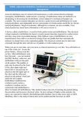
When you get to your units, ask your nurse or clinical instructor go over this. You will find it on
top of the crash cart. Locate the
power button. Always turn this
on first. Most defibrillators only
have the pads – you can monitor
the EKG rhythm, cardiovert, and
defibrillator with just the pads
on the patient. The paddles or
pads may be placed on the front
of the chest upper right and
lateral left or one pad may be
placed on the left upper part of
the chest and the other pad
placed on the patient’s back just
under the left scapula.
Defibrillator multifunction
conductor pads contain a
conductive medium and are
connected to the defibrillator to
allow for hands-off defibrillation. This method reduces the risk of touching the patient during
the procedure and increases electrical safety. In most settings of higher acuity patients, the
nurses are expected to identify shockable/paceable rhythms, but in other areas such as L&D,
Med/Surg, there is the analyze button that can be pushed and this becomes an AED.
1
N4581: Cardioversion, Defibrillation, and Pacemaker Management Overview
, N4581: Adjunctive Modalities: Cardioversion, Defibrillation, and Pacemaker
Management
The energy button increases or decreases the amount of electrical current – this can be between
25 joules up to 360 joules (some only go to 200 joules depending on the manufacturer). The
charge button will “charge” the defibrillator up to that amount of joules when you are ready to
shock the patient. The lightning bolt will deliver the joules. Prior to pushing this button one
needs to make sure no one is touching the patient and the BVM has been removed from the pt.
Anyone touching the patient could also receive the electrical current causing them to develop a
lethal dysrhythmia.
The “synch” button is used in cardioversion. Electrical cardioversion involves the delivery of a
“timed” electrical current synchronized to the patient’s rhythm to terminate a tachycardic
dysrhythmia.
Both defibrillation and cardioversion are used to try to stop the abnormal electrical impulses
traveling through the heart. One major difference between cardioversion and defibrillation is the
timing of the delivery of electrical current. In cardioversion, the delivery of the electrical
current is synchronized with the patient’s electrical events; in defibrillation, the delivery of the
current is immediate and unsynchronized.
Cardioversion will send an electrical impulse at a specific time through the myocardium as an
electrical “reboot” of the conduction system. The defibrillator is set to synchronize with the
ECG on a cardiac monitor so that the electrical impulse discharges during ventricular
depolarization (QRS complex). The synchronization prevents the discharge from occurring
during the vulnerable period of repolarization (T wave), which could result in VT or ventricular
fibrillation. The ECG monitor connected to the external defibrillator usually displays a mark or
line that indicates sensing of a QRS complex. Sometimes the lead and the electrodes must be
changed for the monitor to recognize the patient’s QRS complex. When the synchronizer is on,
no electrical current is delivered if the defibrillator does not identify a QRS complex. Therefore,
it is important to ensure that the patient is connected to the monitor and to select a lead that has
the most appropriate sensing of the QRS. Because there may be a short delay until recognition
of the QRS, the discharge buttons of an external manual defibrillator must be held down until
the shock has been delivered. In most monitors, the synchronization mode must be reactivated if
the initial cardioversion was ineffective and another cardioversion is needed (the device defaults
to unsynchronized defibrillation mode). It is used for tachycardic dysrhythmias electively or in
emergent situations if the patient is starting to decompensate (SOB, angina, hypotension,
diaphoretic).
If the cardioversion is elective and the dysrhythmia such as atrial fibrillation has lasted longer
than 48 hours, anticoagulation for a few weeks before cardioversion may be indicated. Digoxin
is usually withheld for 48 hours before cardioversion to ensure the resumption of sinus rhythm
2
N4581: Cardioversion, Defibrillation, and Pacemaker Management Overview
, N4581: Adjunctive Modalities: Cardioversion, Defibrillation, and Pacemaker
Management
with normal conduction. The patient is instructed not to eat or drink for at least 4 hours before
the procedure. Conductor pads are positioned and the patient will receive moderate sedation IV
as well as an analgesic medication or anesthesia. Respiration is then supported with
supplemental oxygen delivered by a bag-valve mask device if needed with suction equipment
readily available. Although patients rarely require intubation, equipment is nearby in case it is
needed. The amount of voltage used varies from 50 to 360 joules, depending on the
defibrillator’s technology, the type and duration of the dysrhythmia, and the size and
hemodynamic status of the patient. The patient may be cardioverted several times with an
increase in the joules each time. Remember: the default after each shock resets the defibrillator
off synch so the button must be pushed each sequential time. Always confirm the synch is on by
looking at the marking on the screen of the defibrillator monitor.
Indications of a successful response are conversion to sinus rhythm, adequate peripheral pulses,
and adequate blood pressure. Because of the sedation, airway patency must be maintained and
the patient’s state of consciousness assessed. Vital signs and oxygen saturation are monitored
and recorded until the patient is stable and recovered from sedation and analgesic medications
or anesthesia. ECG monitoring is required during and after.
Defibrillation is used in emergency situations as the treatment of choice for ventricular
fibrillation and pulseless VT where the ventricles area chaotically beating and not generating a
pulse with it, the most common cause of abrupt loss of cardiac function and sudden cardiac
death. Defibrillation is not used for patients who are conscious or have a pulse. The energy
setting for the initial and subsequent shocks using a monophasic defibrillator should be set at
360 joules. The energy setting for the initial shock using a biphasic defibrillator may be set at
150 to 200 joules, with the same or an increasing dose with subsequent shocks. The sooner
defibrillation is used, the better the survival rate. Several studies have demonstrated that early
defibrillation performed by lay people in a community setting can increase the survival rate. If
immediate CPR is provided and defibrillation is performed within 5 minutes, more adults in
ventricular fibrillation may survive with intact neurologic function.
Epinephrine is given after initial unsuccessful defibrillation to make it easier to convert the
arrhythmia to a normal rhythm with the next defibrillation. This medication may also increase
cerebral and coronary artery blood flow. Antiarrhythmic medications such as amiodarone,
lidocaine, or magnesium may be given if ventricular arrhythmia persists. This treatment with
continuous CPR, medication administration, and defibrillation continues until a stable rhythm
resumes or until it is determined that the patient cannot be revived.
A pacemaker is an electronic device that provides electrical stimuli to the heart muscle.
Pacemakers are usually used when a patient has a permanent or temporary slower-than-normal
3
N4581: Cardioversion, Defibrillation, and Pacemaker Management Overview
Management
Acute dysrhythmias may be treated with medications or with external electrical therapy
(emergency defibrillation, cardioversion, or pacing). If medications alone are ineffective in
eliminating or decreasing the dysrhythmia, certain adjunctive mechanical therapies are
available. The most common therapies are elective cardioversion and defibrillation for acute
tachydysrhythmia, and implantable devices (pacemakers for bradycardias and ICDs for chronic
tachydysrhythmias). Surgical treatments, although less common, are also available. Your
clinical packet has much more information so be sure to review it.
A device called a defibrillator, is used for both cardioversion and defibrillation. The electrical
voltage required to defibrillate the heart is usually greater than that required for cardioversion
and may cause more myocardial damage. Only biphasic types of defibrillators are now
manufactured; these deliver an electrical charge from one paddle that then automatically
redirects its charge back to the originating paddle. The electrical current may be delivered
externally through the skin with the use of paddles or with conductor pads.
When you get to your units, ask your nurse or clinical instructor go over this. You will find it on
top of the crash cart. Locate the
power button. Always turn this
on first. Most defibrillators only
have the pads – you can monitor
the EKG rhythm, cardiovert, and
defibrillator with just the pads
on the patient. The paddles or
pads may be placed on the front
of the chest upper right and
lateral left or one pad may be
placed on the left upper part of
the chest and the other pad
placed on the patient’s back just
under the left scapula.
Defibrillator multifunction
conductor pads contain a
conductive medium and are
connected to the defibrillator to
allow for hands-off defibrillation. This method reduces the risk of touching the patient during
the procedure and increases electrical safety. In most settings of higher acuity patients, the
nurses are expected to identify shockable/paceable rhythms, but in other areas such as L&D,
Med/Surg, there is the analyze button that can be pushed and this becomes an AED.
1
N4581: Cardioversion, Defibrillation, and Pacemaker Management Overview
, N4581: Adjunctive Modalities: Cardioversion, Defibrillation, and Pacemaker
Management
The energy button increases or decreases the amount of electrical current – this can be between
25 joules up to 360 joules (some only go to 200 joules depending on the manufacturer). The
charge button will “charge” the defibrillator up to that amount of joules when you are ready to
shock the patient. The lightning bolt will deliver the joules. Prior to pushing this button one
needs to make sure no one is touching the patient and the BVM has been removed from the pt.
Anyone touching the patient could also receive the electrical current causing them to develop a
lethal dysrhythmia.
The “synch” button is used in cardioversion. Electrical cardioversion involves the delivery of a
“timed” electrical current synchronized to the patient’s rhythm to terminate a tachycardic
dysrhythmia.
Both defibrillation and cardioversion are used to try to stop the abnormal electrical impulses
traveling through the heart. One major difference between cardioversion and defibrillation is the
timing of the delivery of electrical current. In cardioversion, the delivery of the electrical
current is synchronized with the patient’s electrical events; in defibrillation, the delivery of the
current is immediate and unsynchronized.
Cardioversion will send an electrical impulse at a specific time through the myocardium as an
electrical “reboot” of the conduction system. The defibrillator is set to synchronize with the
ECG on a cardiac monitor so that the electrical impulse discharges during ventricular
depolarization (QRS complex). The synchronization prevents the discharge from occurring
during the vulnerable period of repolarization (T wave), which could result in VT or ventricular
fibrillation. The ECG monitor connected to the external defibrillator usually displays a mark or
line that indicates sensing of a QRS complex. Sometimes the lead and the electrodes must be
changed for the monitor to recognize the patient’s QRS complex. When the synchronizer is on,
no electrical current is delivered if the defibrillator does not identify a QRS complex. Therefore,
it is important to ensure that the patient is connected to the monitor and to select a lead that has
the most appropriate sensing of the QRS. Because there may be a short delay until recognition
of the QRS, the discharge buttons of an external manual defibrillator must be held down until
the shock has been delivered. In most monitors, the synchronization mode must be reactivated if
the initial cardioversion was ineffective and another cardioversion is needed (the device defaults
to unsynchronized defibrillation mode). It is used for tachycardic dysrhythmias electively or in
emergent situations if the patient is starting to decompensate (SOB, angina, hypotension,
diaphoretic).
If the cardioversion is elective and the dysrhythmia such as atrial fibrillation has lasted longer
than 48 hours, anticoagulation for a few weeks before cardioversion may be indicated. Digoxin
is usually withheld for 48 hours before cardioversion to ensure the resumption of sinus rhythm
2
N4581: Cardioversion, Defibrillation, and Pacemaker Management Overview
, N4581: Adjunctive Modalities: Cardioversion, Defibrillation, and Pacemaker
Management
with normal conduction. The patient is instructed not to eat or drink for at least 4 hours before
the procedure. Conductor pads are positioned and the patient will receive moderate sedation IV
as well as an analgesic medication or anesthesia. Respiration is then supported with
supplemental oxygen delivered by a bag-valve mask device if needed with suction equipment
readily available. Although patients rarely require intubation, equipment is nearby in case it is
needed. The amount of voltage used varies from 50 to 360 joules, depending on the
defibrillator’s technology, the type and duration of the dysrhythmia, and the size and
hemodynamic status of the patient. The patient may be cardioverted several times with an
increase in the joules each time. Remember: the default after each shock resets the defibrillator
off synch so the button must be pushed each sequential time. Always confirm the synch is on by
looking at the marking on the screen of the defibrillator monitor.
Indications of a successful response are conversion to sinus rhythm, adequate peripheral pulses,
and adequate blood pressure. Because of the sedation, airway patency must be maintained and
the patient’s state of consciousness assessed. Vital signs and oxygen saturation are monitored
and recorded until the patient is stable and recovered from sedation and analgesic medications
or anesthesia. ECG monitoring is required during and after.
Defibrillation is used in emergency situations as the treatment of choice for ventricular
fibrillation and pulseless VT where the ventricles area chaotically beating and not generating a
pulse with it, the most common cause of abrupt loss of cardiac function and sudden cardiac
death. Defibrillation is not used for patients who are conscious or have a pulse. The energy
setting for the initial and subsequent shocks using a monophasic defibrillator should be set at
360 joules. The energy setting for the initial shock using a biphasic defibrillator may be set at
150 to 200 joules, with the same or an increasing dose with subsequent shocks. The sooner
defibrillation is used, the better the survival rate. Several studies have demonstrated that early
defibrillation performed by lay people in a community setting can increase the survival rate. If
immediate CPR is provided and defibrillation is performed within 5 minutes, more adults in
ventricular fibrillation may survive with intact neurologic function.
Epinephrine is given after initial unsuccessful defibrillation to make it easier to convert the
arrhythmia to a normal rhythm with the next defibrillation. This medication may also increase
cerebral and coronary artery blood flow. Antiarrhythmic medications such as amiodarone,
lidocaine, or magnesium may be given if ventricular arrhythmia persists. This treatment with
continuous CPR, medication administration, and defibrillation continues until a stable rhythm
resumes or until it is determined that the patient cannot be revived.
A pacemaker is an electronic device that provides electrical stimuli to the heart muscle.
Pacemakers are usually used when a patient has a permanent or temporary slower-than-normal
3
N4581: Cardioversion, Defibrillation, and Pacemaker Management Overview