
Structure and Function of the CV System
The circulatory system includes the heart and blood vessels. The heart’s job is to pump, providing continuous
rhythmic contractions. The arterial system distributes oxygenated blood to tissues. The venous system collects
deoxygenated blood & returns it to the heart. The capillaries are where the exchange of gasses, nutrients &
wastes takes place
Functions of the cardiovascular system → transportation and delivery of oxygen & nutrients. Carries waste
products for elimination. Circulates electrolytes & hormones, and regulates body temperature
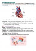
Heart anatomy → a hollow muscular organ in the thorax between the lungs (lungs act as a cushion). 4
chambers, 2 atriums, and 2 ventricles. Two separate pumps (left and right sides)
● Right side → receives blood from the body & sends it to the lungs (pulmonary)
● Left side → receives oxygenated blood from the lungs and sends it to the body (systemic) – a little
more posterior than the right side; more protected
Pericardium, myocardium, endocardium, and heart valves
Atria → thin-walled reservoirs for blood
Ventricles → thick-walled for pumping blood out of the heart
Pericardium → loose fitting sac around the heart, holding it in place in a fixed position; provides physical
protection & a barrier to infection (not necessary for heart function)
Myocardium → muscular portion forms the wall of the atria & ventricles
Endocardium → thin, 3 layered membrane lining the inside of the heart; forms the surface of the valves
One-way valves → AV valves & semilunar valves are pressure valves to make sure there is only a one-way
flow
● AV valves = tricuspid and mitral valves
● Semilunar valve = aortic and pulmonary valves
Fibrous skeleton → provides structural support & isolating force for the electrical impulse
,Deoxygenated blood comes into the R atrium from the superior & inferior vena cava → tricuspid valve → into
the R ventricle → pulmonary valve into the lungs for oxygenation via the pulmonary arteries → oxygenated
blood comes into the L atrium via the pulmonary veins → through the mitral valve during diastole → into the L
ventricle → through the aortic valve → pumped out to the body by the aorta
Pulmonary Circulation (R ♡) Systemic Circulation (L ♡)
Moves blood through the lungs & creates a link with Supplies all other tissues of the body
the gas exchange function of the respiratory system ● L heart
● R heart ● Aorta & its branches
● Pulmonary artery ● Capillaries supplying the brain & peripheral
● Capillaries & veins tissues
● Systemic venous system & vena cava
How does blood flow through blood vessels? → from higher pressure to lower pressure in the vessels,
heart, and valves
● Blood flow (F) = the pressure difference / the resistance to flow
● Resistance = opposition of flow caused by friction between the moving blood & the stationary vessel
wall
Peripheral vascular resistance (PVR) & Systemic vascular resistance (SVR) = collective resistance of all
vessels in peripheral circulation
Blood flow = change in pressure x 𝞹 x radius to the 4th power OVER 8 x length of the vessel x the viscosity of
blood
The radius has a big effect on vessels; a smaller radius = < blood flow → Example: a vasodilator will ↑ the
radius of the vessel = ↑ blood flow = ↑ oxygenation
, Radius (1mm) to the 4th power = 1 mL/min
Radius (2mm) to the 4th power = 16 mL/min
The dynamic/changeable components of Poiseuille’s equation are:
● The viscosity of blood – can change a little bit as Hct goes up
● Radius of vessels is always changing – can constrict or dilate (radius has the biggest impact on blood
flow)
Cardiac Cycle → rhythmic pumping action of the heart
Systole = ventricles are contracting
● Semilunar valves (aortic & pulmonary valves) are open
● AV valves (tricuspid and mitral valves) are closed during contraction
Diastole = ventricles are relaxed and filling with blood → AV valves are open, letting blood pour into the
ventricles from the atria
Measuring the efficiency of the heart
End-diastolic volume (EDV) = amount of blood in the ventricles at the end of filling time (diastole)
End-systolic volume (ESV) = amount of blood remaining in the ventricles at the end of systole (after ejection)
Stroke volume (SV) = EDV - ESV
Ejection fraction (EF) = SV / EDV (Normal EF is 55-75%)
, Top picture → Events on the left side of the heart showing changes in aortic pressure, left ventricular pressure,
atrial pressure, left ventricular volume, the ECG, and heart sounds during the cardiac cycle
Bottom picture → AV & semilunar valves during contraction and ventricular ejection, relaxation & ventricular
filling, atrial contraction
Preload, afterload, cardiac contractility, heart rate, and cardiac output
Cardiac Output (CO) = amount of blood the heart pumps out into the circulatory system each minute
● CO = SV x HR
● Stroke volume = amount of blood pumped with each beat
● Heart rate = number of time heart beats each minute
What is CO dependent on?
1. Preload = VOLUME of blood (end-diastolic pressure) = filling & stretching
2. Afterload = PRESSURE the ventricles pump against to get blood out to the body
3. Cardiac contractility (calcium is really important in this)
4. HR
CO increases as HR increases to a certain point…then as HR continues to increase that means there is less
time for diastole = less time for the heart to fill = less blood will be pumped out = decreased CO
Cardiac reserve → maximum % of the increase in CO achieved above normal resting level
Frank-Starling Mechanism → the > the volume of blood in the heart before contraction, the > the volume of
blood ejected from the heart (from increased contractility from EDV stretch)
● The ventricles adjust their pumping ability based on volume
Vascular system → delivers oxygen & nutrients and removes waste from the tissues. Made up of arteries &
arterioles, capillaries, venules & veins
Arteries → thick-walled vessels with large amounts of elastic fibers that stretch during cardiac systole & recoil
during diastole
Arterioles → resistance vessels for the circulatory system; act as control valves where blood is released as it
moves into the capillaries
Venules → collect blood from the capillaries
Veins → transport blood back to the heart; capable of enlarging & storing large quantities of blood
The circulatory system includes the heart and blood vessels. The heart’s job is to pump, providing continuous
rhythmic contractions. The arterial system distributes oxygenated blood to tissues. The venous system collects
deoxygenated blood & returns it to the heart. The capillaries are where the exchange of gasses, nutrients &
wastes takes place
Functions of the cardiovascular system → transportation and delivery of oxygen & nutrients. Carries waste
products for elimination. Circulates electrolytes & hormones, and regulates body temperature
Heart anatomy → a hollow muscular organ in the thorax between the lungs (lungs act as a cushion). 4
chambers, 2 atriums, and 2 ventricles. Two separate pumps (left and right sides)
● Right side → receives blood from the body & sends it to the lungs (pulmonary)
● Left side → receives oxygenated blood from the lungs and sends it to the body (systemic) – a little
more posterior than the right side; more protected
Pericardium, myocardium, endocardium, and heart valves
Atria → thin-walled reservoirs for blood
Ventricles → thick-walled for pumping blood out of the heart
Pericardium → loose fitting sac around the heart, holding it in place in a fixed position; provides physical
protection & a barrier to infection (not necessary for heart function)
Myocardium → muscular portion forms the wall of the atria & ventricles
Endocardium → thin, 3 layered membrane lining the inside of the heart; forms the surface of the valves
One-way valves → AV valves & semilunar valves are pressure valves to make sure there is only a one-way
flow
● AV valves = tricuspid and mitral valves
● Semilunar valve = aortic and pulmonary valves
Fibrous skeleton → provides structural support & isolating force for the electrical impulse
,Deoxygenated blood comes into the R atrium from the superior & inferior vena cava → tricuspid valve → into
the R ventricle → pulmonary valve into the lungs for oxygenation via the pulmonary arteries → oxygenated
blood comes into the L atrium via the pulmonary veins → through the mitral valve during diastole → into the L
ventricle → through the aortic valve → pumped out to the body by the aorta
Pulmonary Circulation (R ♡) Systemic Circulation (L ♡)
Moves blood through the lungs & creates a link with Supplies all other tissues of the body
the gas exchange function of the respiratory system ● L heart
● R heart ● Aorta & its branches
● Pulmonary artery ● Capillaries supplying the brain & peripheral
● Capillaries & veins tissues
● Systemic venous system & vena cava
How does blood flow through blood vessels? → from higher pressure to lower pressure in the vessels,
heart, and valves
● Blood flow (F) = the pressure difference / the resistance to flow
● Resistance = opposition of flow caused by friction between the moving blood & the stationary vessel
wall
Peripheral vascular resistance (PVR) & Systemic vascular resistance (SVR) = collective resistance of all
vessels in peripheral circulation
Blood flow = change in pressure x 𝞹 x radius to the 4th power OVER 8 x length of the vessel x the viscosity of
blood
The radius has a big effect on vessels; a smaller radius = < blood flow → Example: a vasodilator will ↑ the
radius of the vessel = ↑ blood flow = ↑ oxygenation
, Radius (1mm) to the 4th power = 1 mL/min
Radius (2mm) to the 4th power = 16 mL/min
The dynamic/changeable components of Poiseuille’s equation are:
● The viscosity of blood – can change a little bit as Hct goes up
● Radius of vessels is always changing – can constrict or dilate (radius has the biggest impact on blood
flow)
Cardiac Cycle → rhythmic pumping action of the heart
Systole = ventricles are contracting
● Semilunar valves (aortic & pulmonary valves) are open
● AV valves (tricuspid and mitral valves) are closed during contraction
Diastole = ventricles are relaxed and filling with blood → AV valves are open, letting blood pour into the
ventricles from the atria
Measuring the efficiency of the heart
End-diastolic volume (EDV) = amount of blood in the ventricles at the end of filling time (diastole)
End-systolic volume (ESV) = amount of blood remaining in the ventricles at the end of systole (after ejection)
Stroke volume (SV) = EDV - ESV
Ejection fraction (EF) = SV / EDV (Normal EF is 55-75%)
, Top picture → Events on the left side of the heart showing changes in aortic pressure, left ventricular pressure,
atrial pressure, left ventricular volume, the ECG, and heart sounds during the cardiac cycle
Bottom picture → AV & semilunar valves during contraction and ventricular ejection, relaxation & ventricular
filling, atrial contraction
Preload, afterload, cardiac contractility, heart rate, and cardiac output
Cardiac Output (CO) = amount of blood the heart pumps out into the circulatory system each minute
● CO = SV x HR
● Stroke volume = amount of blood pumped with each beat
● Heart rate = number of time heart beats each minute
What is CO dependent on?
1. Preload = VOLUME of blood (end-diastolic pressure) = filling & stretching
2. Afterload = PRESSURE the ventricles pump against to get blood out to the body
3. Cardiac contractility (calcium is really important in this)
4. HR
CO increases as HR increases to a certain point…then as HR continues to increase that means there is less
time for diastole = less time for the heart to fill = less blood will be pumped out = decreased CO
Cardiac reserve → maximum % of the increase in CO achieved above normal resting level
Frank-Starling Mechanism → the > the volume of blood in the heart before contraction, the > the volume of
blood ejected from the heart (from increased contractility from EDV stretch)
● The ventricles adjust their pumping ability based on volume
Vascular system → delivers oxygen & nutrients and removes waste from the tissues. Made up of arteries &
arterioles, capillaries, venules & veins
Arteries → thick-walled vessels with large amounts of elastic fibers that stretch during cardiac systole & recoil
during diastole
Arterioles → resistance vessels for the circulatory system; act as control valves where blood is released as it
moves into the capillaries
Venules → collect blood from the capillaries
Veins → transport blood back to the heart; capable of enlarging & storing large quantities of blood


