
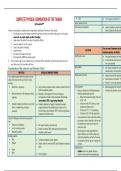
COMPLETE PHYSICAL EXAMINATION OF THE THORAX § Rate ⇒ 14- 20 cycles/ min and I:E of 1: 2
seffcausapinmd™ Lumps, lesions and scars
Extra- thoracic observations ⇒ cyanosis, clubbing in patients wit
There are several points to remember prior to beginning the examination of the thorax. These include: edema in patients with pleural flu
1. Even though you may have already recorded the respiratory rate when you took the vital signs, it is wise to again
observe the rate, rhythm, depth, and effort of breathing.
2. Always inspect the patient for any signs of respiratory difficulty.
3. Assess the patient’s color for cyanosis
4. Listen to the patient’s breathing Focus on areas of tenderness and abn
5. Inspect the neck. PALPATION
respiratory expansion, and fremitus
6. Also observe the shape of the chest.
7. Most importantly, DRAPE the patient properly. Identify tender areas ⇒ Intercostal tenderness over inflam
∇ These can also be part of your inspection as you go through with the examination, but the book says these are part of ⇒ Local tenderness from trauma ma
your initial survey of the respiration and thorax. from costochondritis
Examination of the Anterior and Posterior Chest Abnormalities ⇒ Like crepitus (sensation of crackl
subcutaneous air or pneumothor
INSPECTION RATIONALE/ SIGNIFICANT FINDINGS
Diaphragmatic excursion ⇒ Unequal movement à asymmetr
From a midline position behind the patient, note the
shape of the chest and the way in which it moves, ⇒ Minute amount of movement à
including: ⇒ Normal: 5- 6cm on full inspiration
§ Deformities or asymmetry ⇒ Such as pectus excavatum, pectus carinatum, and barrel chest in Chest expansion ⇒ Unilateral lagging from chronic fib
COPD and asthmatic patients ◦ Place your thumbs on the following landmarks restrictive lung disorders and uni
§ Abnormal retraction of the interspaces during ⇒ Retraction is most apparent in the lower interspaces. following cephalocaudal approach:
inspiration Supraclavicular retraction is often associated. Retraction in a. Below the sternoclavicular joint at the level
severe asthma, COPD, or upper airway obstruction. of the 1st rib
§ Impaired respiratory movement on one or both ⇒ Unilateral impairment or lagging of respiratory movement b. At the level of the T4/T5 with your hand
sides or a unilateral lag (or delay) in suggests disease of the underlying lung or pleura such as grasping the axillary portion
movement. chronic fibrosis, pleural effusion, COPD, restrictive lung diseases, c. At the level of T11- T12 or diaphragm
unilateral bronchial obstruction. d. At the level of the 10th ribs (posteriorly)
Inspect the following in both anterior and posterior ◦ As you position your hands, slide them medially
view. just enough to raise a loose fold of skin on each
§ Patient’s level of distress. side between your thumb and the spine (in
⇒ SCM, upper trapezius, pectoralis major
§ Use of respiratory muscles posterior portion)
⇒ Patients w/ asthma/ COPD, sitting and leaning forward with
§ Respiratory positions ◦ Ask the patient to inhale deeply. Watch the
shoulders arched forward.
distance between your thumbs as they move
Breathing patterns: apart during inspiration, and feel for the range
§ Rate ⇒ Tachypnea, bradypnea, apnea, dyspnea and symmetry of the rib cage as it expands and
§ Rhythym ⇒ Cheyne- stokes in uremia and CHF, biots breathing in respiratory contracts.
failure Tactile fremitus
§ Depth
- Fremitus refers to the palpable vibrations
§ Effort ⇒ Such as dyspnea transmit- ted through the bronchopulmonary
, tree to the chest wall when the patient speaks PATHOLOGIC EXAMPLES:
Procedure: § Dullness- pleural effusion, atele
◦ To detect fremitus, use either the ball (the consolidation
bony part of the palm at the base of the § Hyper- resonance- emphysema
fingers) or the ulnar surface of your hand § Tympany- very large pneumoth
to optimize the vibratory sensitivity of the NORMAL AREAS OF DULLNESS
bones in your hand. Ask the patient to § Cardiac dullness- 3- 5th ICS, L
repeat the words “ninety-nine” or “one- § Liver span- 4- 6th ICS, Right M
one-one.” If fremitus is faint, ask the ⇒ Decreased tactile fremitus in pneumothorax, pleural effusion, § Splenic dullness- 9- 11th rib,
patient to speak more loudly or in a deeper and thick chest § Level of diaphragm- 5- 6 cm
voice. ⇒ Increased tactile fremitus in mass, consolidation, solid organs Locations for percussion
and thin chest
Percussion of the chest sets the chest wall and underlying tissues into
motion, producing audible sound and palpable vibrations. Percussion
helps you establish whether the underlying tissues are air-filled, fluid-
PERCUSSION
filled, or solid. It penetrates only about 5 cm to 7 cm into the chest,
however, and therefore will not help you to detect deep-seated
lesions.
• Hyperextend the middle finger of your left hand,
known as the pleximeter finger.
• Press its distal interphalangeal joint firmly on the Auscultation of the lungs is the most
AUSCULTATION
surface to be percussed. for assessing air flow through the tra
• Avoid surface contact by any other part of the Auscultation involves:
hand, because this dampens out vibrations. (1) listening to the sounds generated by breathing
• Note that the thumb, 2nd, 4th, and 5th fingers (2) listening for any adventitious (added) sounds, and
are not touching the chest. (3) if abnormalities are suspected, listening to the
• Position your right forearm quite close to the sounds of the patient’s spoken or whispered voice as
surface, with the hand cocked upward. The they are transmitted through the chest wall.
middle finger should be partially flexed, relaxed,
and poised to strike.
Learn to identify five percussion notes.
, Adventitious Breath Sounds
§ Crackles may be due to abnormalities of the
lungs (pneumonia, fibrosis, early congestive
heart failure) or of the air- ways (bronchitis,
bronchiectasis).
§ Wheezes suggest narrowed airways, as in
asthma, COPD, or bronchitis.
§ Rhonchi suggest secretions in large airways.
§ Fine late inspiratory crackles that persist from
breath to breath suggest abnormal lung tissue.
Transmitted Voice Sounds
§ Bronchophony- Ask the patient to say “ninety- ⇒ Louder, clearer voice sounds are called bronchophony.
nine.” Normally the sounds transmitted through
the chest wall are muffled and indistinct.
§ Egophony- Ask the patient to say “ee.” You will ⇒ When “ee” is heard as “ay,” an E-to-A change (egophony) is
normally hear a muffled long E sound. present, as in lobar consolidation from pneumonia. The quality
sounds nasal.
§ Pectoriloquy- Ask the patient to whisper “ninety- ⇒ Louder, clearer whispered sounds are called whispered
nine” or “one-two-three.” The whispered voice pectoriloquy.
is normally heard faintly and indistinctly, if at all.
SPECIAL TECHNIQUES IMPORTANT POINTS TO CONSIDER
Clinical Assessment of Pulmonary Function - assess the complaint of breathlessness in an ambulatory
patient is to walk with the patient down the hall or climb one
flight of stairs. Observe the rate, effort, and sound of the
patient’s breathing
Forced Expiratory Time - Ask the patient to take a deep breath in and then breathe out
as quickly and completely as possible with mouth open.
Listen over the trachea with the diaphragm of a stethoscope
and time the audible expiration. Try to get three consistent
readings, allowing a short rest between efforts if necessary.
Identification of a Fractured Rib - An increase in the local pain (distant from your hands)
- By anteroposterior compression of the chest, you suggests rib fracture rather than just soft tissue injury.
can help to distinguish a fracture from soft-tissue
injury. With one hand on the sternum and the other
on the thoracic spine, squeeze the chest. Is this
painful, and where?
seffcausapinmd™ Lumps, lesions and scars
Extra- thoracic observations ⇒ cyanosis, clubbing in patients wit
There are several points to remember prior to beginning the examination of the thorax. These include: edema in patients with pleural flu
1. Even though you may have already recorded the respiratory rate when you took the vital signs, it is wise to again
observe the rate, rhythm, depth, and effort of breathing.
2. Always inspect the patient for any signs of respiratory difficulty.
3. Assess the patient’s color for cyanosis
4. Listen to the patient’s breathing Focus on areas of tenderness and abn
5. Inspect the neck. PALPATION
respiratory expansion, and fremitus
6. Also observe the shape of the chest.
7. Most importantly, DRAPE the patient properly. Identify tender areas ⇒ Intercostal tenderness over inflam
∇ These can also be part of your inspection as you go through with the examination, but the book says these are part of ⇒ Local tenderness from trauma ma
your initial survey of the respiration and thorax. from costochondritis
Examination of the Anterior and Posterior Chest Abnormalities ⇒ Like crepitus (sensation of crackl
subcutaneous air or pneumothor
INSPECTION RATIONALE/ SIGNIFICANT FINDINGS
Diaphragmatic excursion ⇒ Unequal movement à asymmetr
From a midline position behind the patient, note the
shape of the chest and the way in which it moves, ⇒ Minute amount of movement à
including: ⇒ Normal: 5- 6cm on full inspiration
§ Deformities or asymmetry ⇒ Such as pectus excavatum, pectus carinatum, and barrel chest in Chest expansion ⇒ Unilateral lagging from chronic fib
COPD and asthmatic patients ◦ Place your thumbs on the following landmarks restrictive lung disorders and uni
§ Abnormal retraction of the interspaces during ⇒ Retraction is most apparent in the lower interspaces. following cephalocaudal approach:
inspiration Supraclavicular retraction is often associated. Retraction in a. Below the sternoclavicular joint at the level
severe asthma, COPD, or upper airway obstruction. of the 1st rib
§ Impaired respiratory movement on one or both ⇒ Unilateral impairment or lagging of respiratory movement b. At the level of the T4/T5 with your hand
sides or a unilateral lag (or delay) in suggests disease of the underlying lung or pleura such as grasping the axillary portion
movement. chronic fibrosis, pleural effusion, COPD, restrictive lung diseases, c. At the level of T11- T12 or diaphragm
unilateral bronchial obstruction. d. At the level of the 10th ribs (posteriorly)
Inspect the following in both anterior and posterior ◦ As you position your hands, slide them medially
view. just enough to raise a loose fold of skin on each
§ Patient’s level of distress. side between your thumb and the spine (in
⇒ SCM, upper trapezius, pectoralis major
§ Use of respiratory muscles posterior portion)
⇒ Patients w/ asthma/ COPD, sitting and leaning forward with
§ Respiratory positions ◦ Ask the patient to inhale deeply. Watch the
shoulders arched forward.
distance between your thumbs as they move
Breathing patterns: apart during inspiration, and feel for the range
§ Rate ⇒ Tachypnea, bradypnea, apnea, dyspnea and symmetry of the rib cage as it expands and
§ Rhythym ⇒ Cheyne- stokes in uremia and CHF, biots breathing in respiratory contracts.
failure Tactile fremitus
§ Depth
- Fremitus refers to the palpable vibrations
§ Effort ⇒ Such as dyspnea transmit- ted through the bronchopulmonary
, tree to the chest wall when the patient speaks PATHOLOGIC EXAMPLES:
Procedure: § Dullness- pleural effusion, atele
◦ To detect fremitus, use either the ball (the consolidation
bony part of the palm at the base of the § Hyper- resonance- emphysema
fingers) or the ulnar surface of your hand § Tympany- very large pneumoth
to optimize the vibratory sensitivity of the NORMAL AREAS OF DULLNESS
bones in your hand. Ask the patient to § Cardiac dullness- 3- 5th ICS, L
repeat the words “ninety-nine” or “one- § Liver span- 4- 6th ICS, Right M
one-one.” If fremitus is faint, ask the ⇒ Decreased tactile fremitus in pneumothorax, pleural effusion, § Splenic dullness- 9- 11th rib,
patient to speak more loudly or in a deeper and thick chest § Level of diaphragm- 5- 6 cm
voice. ⇒ Increased tactile fremitus in mass, consolidation, solid organs Locations for percussion
and thin chest
Percussion of the chest sets the chest wall and underlying tissues into
motion, producing audible sound and palpable vibrations. Percussion
helps you establish whether the underlying tissues are air-filled, fluid-
PERCUSSION
filled, or solid. It penetrates only about 5 cm to 7 cm into the chest,
however, and therefore will not help you to detect deep-seated
lesions.
• Hyperextend the middle finger of your left hand,
known as the pleximeter finger.
• Press its distal interphalangeal joint firmly on the Auscultation of the lungs is the most
AUSCULTATION
surface to be percussed. for assessing air flow through the tra
• Avoid surface contact by any other part of the Auscultation involves:
hand, because this dampens out vibrations. (1) listening to the sounds generated by breathing
• Note that the thumb, 2nd, 4th, and 5th fingers (2) listening for any adventitious (added) sounds, and
are not touching the chest. (3) if abnormalities are suspected, listening to the
• Position your right forearm quite close to the sounds of the patient’s spoken or whispered voice as
surface, with the hand cocked upward. The they are transmitted through the chest wall.
middle finger should be partially flexed, relaxed,
and poised to strike.
Learn to identify five percussion notes.
, Adventitious Breath Sounds
§ Crackles may be due to abnormalities of the
lungs (pneumonia, fibrosis, early congestive
heart failure) or of the air- ways (bronchitis,
bronchiectasis).
§ Wheezes suggest narrowed airways, as in
asthma, COPD, or bronchitis.
§ Rhonchi suggest secretions in large airways.
§ Fine late inspiratory crackles that persist from
breath to breath suggest abnormal lung tissue.
Transmitted Voice Sounds
§ Bronchophony- Ask the patient to say “ninety- ⇒ Louder, clearer voice sounds are called bronchophony.
nine.” Normally the sounds transmitted through
the chest wall are muffled and indistinct.
§ Egophony- Ask the patient to say “ee.” You will ⇒ When “ee” is heard as “ay,” an E-to-A change (egophony) is
normally hear a muffled long E sound. present, as in lobar consolidation from pneumonia. The quality
sounds nasal.
§ Pectoriloquy- Ask the patient to whisper “ninety- ⇒ Louder, clearer whispered sounds are called whispered
nine” or “one-two-three.” The whispered voice pectoriloquy.
is normally heard faintly and indistinctly, if at all.
SPECIAL TECHNIQUES IMPORTANT POINTS TO CONSIDER
Clinical Assessment of Pulmonary Function - assess the complaint of breathlessness in an ambulatory
patient is to walk with the patient down the hall or climb one
flight of stairs. Observe the rate, effort, and sound of the
patient’s breathing
Forced Expiratory Time - Ask the patient to take a deep breath in and then breathe out
as quickly and completely as possible with mouth open.
Listen over the trachea with the diaphragm of a stethoscope
and time the audible expiration. Try to get three consistent
readings, allowing a short rest between efforts if necessary.
Identification of a Fractured Rib - An increase in the local pain (distant from your hands)
- By anteroposterior compression of the chest, you suggests rib fracture rather than just soft tissue injury.
can help to distinguish a fracture from soft-tissue
injury. With one hand on the sternum and the other
on the thoracic spine, squeeze the chest. Is this
painful, and where?


