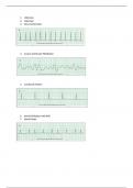
● Skeletal muscle is responsible for voluntary movement of the human body.
Skeletal muscles are made up of muscle fibers with connective tissue wrappings.
● The dense, fibrous connective tissue surrounding the entire muscle is called the
epimysium. At the ends of the muscle, the epimysium is continuous with the
tendons and periosteum
● Thin projections of connective tissue called perimysium extend from the
epimysium into the muscle to surround bundles of muscle fibers known as
fascicles. Within each fascicle, loose connective tissue called endomysium
separates individual muscle fibers.
● A muscle fiber consists of a single, elongated cell surrounded by a plasma
membrane, known as the sarcolemma. Each muscle fiber contains muscle nuclei
that are found just beneath the sarcolemma.
● Threadlike structures called myofibrils extend the length of the fiber and dominate
its interior. Myofibrils, which are unique to muscle, are composed of two kinds of
protein filaments- actin (or thin) myofilaments and myosin (or thick)
myofilaments.
● The actin and myosin myofilaments are organized in an orderly contractile unit
called a sarcomere. The sliding of these filaments along each other shortened
sarcomeres result in muscle contraction and, ultimately, movement of the skeleton.
● Skeletal muscles are voluntary, controlled consciously by the nervous system, and
are made up of fascicles, which are bundles of muscle fibers surrounded by
connective tissue, blood vessels, and nerves.
● Motor nerve axons supply one or more skeletal muscle fibers constituting a motor
unit. Inside a muscle fiber, thread-like structures called myofibrils are organized
into contractile units, or sarcomeres, reflecting the striations characteristics of
skeletal muscle. Each sarcomere is made up of thick and thin myofilaments,
strands of proteins called myosin and actin.
● Myosin forms thick filaments. Actin forms thin filaments. The protruding heads of
myosin filaments form cross bridges that bind to actin filaments and ATP, an
energy transport molecule.
● Thin filaments contain a sequence of actin molecules with myosin binding sites.
● At rest, actin molecules bind tropomyosin and troponin, two contractile proteins
that inhibit contraction.
● Skeletal muscle contraction begins at the muscle fibers’ plasma membrane, or
sarcolemma. With invaginations called transverse, or T-tubules, that surround each
myofibril and open to the extracellular space.