

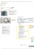
Receptors- Eye
Thursday, 11 July 2024 16:55
3 layers of internal structure: -outer fibrous layer -middle vascular layer -inner light
sensitive layer
-outer fibrous layer: sclera, cornea
Sclera: thick layer of white inelastic connective tissue that forms the outer layer of
the eyeball and extends to the cornea at the front
FUNCTION: to protect the internal parts of the eyeball, helps to maintain spherical
shape of eye, for muscle attachment
Cornea: transparent front part of the sclera, more convex than rest of eyeball
FUNCTION: to let light rays through to inner layer of eye, causing refraction of
incoming light rays
-middle vascular layer: choroid, ciliary body, suspensory ligaments, lens, iris, pupil
Choroid: thin darkly pigmented vascular layer
FUNCTION: to absorb excess light rays to prevent internal reflection which may cause
blurred images. Blood vessels supply oxygen and nutrients to cells
Ciliary body: thickening of front part of choroid. Consists of involuntary muscles
[ciliary muscles]. Attached to lens by suspensory ligaments.
FUNCTION: to relax and contract to change curvature of lens during accommodation
Iris: round coloured structure situated in centre of the eye. Contains pigments and
two sets of involuntary muscles . Radial and circular muscles found here. Opening in
middle of iris is the pupil.
FUNCTION: to regulate the amount of light entering the eye by controlling the size of
the pupil
Lens: rubber, elastic and transparent. Bio convex structure that changes shape.
FUNCTION: changes shape to refract light rays from near and far objects so that a
clear image on retina is formed
OPTIC NERVE: nerve fibres from rods and cones form a synapse with bipolar neuron.
This forms a synapse with optic neurons. Optic neurons join together to form the
optic nerve
Life Science Page 1
Thursday, 11 July 2024 16:55
3 layers of internal structure: -outer fibrous layer -middle vascular layer -inner light
sensitive layer
-outer fibrous layer: sclera, cornea
Sclera: thick layer of white inelastic connective tissue that forms the outer layer of
the eyeball and extends to the cornea at the front
FUNCTION: to protect the internal parts of the eyeball, helps to maintain spherical
shape of eye, for muscle attachment
Cornea: transparent front part of the sclera, more convex than rest of eyeball
FUNCTION: to let light rays through to inner layer of eye, causing refraction of
incoming light rays
-middle vascular layer: choroid, ciliary body, suspensory ligaments, lens, iris, pupil
Choroid: thin darkly pigmented vascular layer
FUNCTION: to absorb excess light rays to prevent internal reflection which may cause
blurred images. Blood vessels supply oxygen and nutrients to cells
Ciliary body: thickening of front part of choroid. Consists of involuntary muscles
[ciliary muscles]. Attached to lens by suspensory ligaments.
FUNCTION: to relax and contract to change curvature of lens during accommodation
Iris: round coloured structure situated in centre of the eye. Contains pigments and
two sets of involuntary muscles . Radial and circular muscles found here. Opening in
middle of iris is the pupil.
FUNCTION: to regulate the amount of light entering the eye by controlling the size of
the pupil
Lens: rubber, elastic and transparent. Bio convex structure that changes shape.
FUNCTION: changes shape to refract light rays from near and far objects so that a
clear image on retina is formed
OPTIC NERVE: nerve fibres from rods and cones form a synapse with bipolar neuron.
This forms a synapse with optic neurons. Optic neurons join together to form the
optic nerve
Life Science Page 1



