
UCI BioSci D170 Epithelial and Connective Tissues Williams
Reading: Chapter 4
What are the four basic types of tissue in the body?
Epithelial, connective, muscle, nervous.
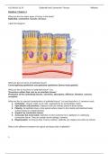
Label this diagram:
What are the two forms of epithelial tissue?
Covering/lining epithelium and glandular epithelium (forms body glands).
What are the six functions of epithelial tissue? List.
*Functions reflect their role as an interface tissue.*
Protection of the underlying tissues, secretion, absorption, diffusion, filtration, sensory
reception.
What are the six special characteristics of epithelial tissue? List and describe in 1 sentence each.
1. Cellularity. Tissues made up of cells, separated by an extracellular matrix.
2. Specialized cell junctions. Adjacent cells are directly joined at these points.
3. Polarity. All epithelia have a free apical surface (open to the inside) and attached basal
surface (lying on a basal lamina).
4. Support by connective tissue.
5. Avascular but innervated. Epithelia receive nutrients from capillaries in underlying
connective tissue. They do contain nerve endings, however.
6. Regeneration. As long as they receive nutrition, epithelia can replace lost cells via mitosis.
What is the difference between the apical and basal side of epithelia?
1
, Apical region is the side that is facing the inner surface of the body (“border an open space”);
usually contains cilia, microvilli. Basal region is the side that sits on the underlying
connective tissue-- the basal lamina.
What are two ways that epithelia are classified?
1) The number of cell layers and 2) the cells’ shape.
Complete the following table to compare and contrast the types of epithelia in the human body.
Epithelial # of
Type(s) of cells Function Examples
type layers
Single layer of
flattened cells w/ Kidney glomeruli, alveoli,
Simple Diffusion and
1 disc-shaped nuclei heart lining, blood vessels,
squamous filtration
and sparse lymphatic vessels, serosae.
cytoplasm.
Single layer of cube- Kidney tubules, ducts and
Simple Secretion and
1 like cells w/ spherical secretory portions of small
cuboidal absorption
central nuclei. glands, ovary surface.
Secretion and
Single layer of tall Line digestive tract,
absorption;
cells w/ oval-like gallbladder, excretory ducts
Simple ciliated types
1 nuclei. Some bear of glands. Ciliated type lines
columnar propel mucus or
microvilli, cilia, or small bronchi, uterine tubes/
are reproductive
goblet cells. some regions of the uterus.
cells.
Ciliated type lines the
Single layer of cells
Pseudostrat trachea and upper
of differing heights. Secretion (i.e.
ified 1 respiratory tract. Non-
Many bear cilia and propels mucus)
columnar ciliated types in male- and
goblet cells.
large gland-ducts.
Several layers of
flattened surface
cells and
cuboidal/columnar
basal cells. Protection of
Line esophagus, mouth,
Stratified Keratinized type: underlying
>1 vagina. Keratinized type
squamous surface cells are tissues subject
form the epidermis.
dead/full of keratin. to abrasion.
Basal cells are
mitotically active and
produce new surface
cells.
Largest ducts of sweat,
Stratified ~2 layers of cube-like
>1 Protection mammary, and salivary
cuboidal cells.
glands.
Basal cells are Rare in humans. Small
Stratified usually cuboidal; Protection and amounts in male urethra and
>1
columnar surface cells are secretion in large ducts of some
columnar. glands.
2
Reading: Chapter 4
What are the four basic types of tissue in the body?
Epithelial, connective, muscle, nervous.
Label this diagram:
What are the two forms of epithelial tissue?
Covering/lining epithelium and glandular epithelium (forms body glands).
What are the six functions of epithelial tissue? List.
*Functions reflect their role as an interface tissue.*
Protection of the underlying tissues, secretion, absorption, diffusion, filtration, sensory
reception.
What are the six special characteristics of epithelial tissue? List and describe in 1 sentence each.
1. Cellularity. Tissues made up of cells, separated by an extracellular matrix.
2. Specialized cell junctions. Adjacent cells are directly joined at these points.
3. Polarity. All epithelia have a free apical surface (open to the inside) and attached basal
surface (lying on a basal lamina).
4. Support by connective tissue.
5. Avascular but innervated. Epithelia receive nutrients from capillaries in underlying
connective tissue. They do contain nerve endings, however.
6. Regeneration. As long as they receive nutrition, epithelia can replace lost cells via mitosis.
What is the difference between the apical and basal side of epithelia?
1
, Apical region is the side that is facing the inner surface of the body (“border an open space”);
usually contains cilia, microvilli. Basal region is the side that sits on the underlying
connective tissue-- the basal lamina.
What are two ways that epithelia are classified?
1) The number of cell layers and 2) the cells’ shape.
Complete the following table to compare and contrast the types of epithelia in the human body.
Epithelial # of
Type(s) of cells Function Examples
type layers
Single layer of
flattened cells w/ Kidney glomeruli, alveoli,
Simple Diffusion and
1 disc-shaped nuclei heart lining, blood vessels,
squamous filtration
and sparse lymphatic vessels, serosae.
cytoplasm.
Single layer of cube- Kidney tubules, ducts and
Simple Secretion and
1 like cells w/ spherical secretory portions of small
cuboidal absorption
central nuclei. glands, ovary surface.
Secretion and
Single layer of tall Line digestive tract,
absorption;
cells w/ oval-like gallbladder, excretory ducts
Simple ciliated types
1 nuclei. Some bear of glands. Ciliated type lines
columnar propel mucus or
microvilli, cilia, or small bronchi, uterine tubes/
are reproductive
goblet cells. some regions of the uterus.
cells.
Ciliated type lines the
Single layer of cells
Pseudostrat trachea and upper
of differing heights. Secretion (i.e.
ified 1 respiratory tract. Non-
Many bear cilia and propels mucus)
columnar ciliated types in male- and
goblet cells.
large gland-ducts.
Several layers of
flattened surface
cells and
cuboidal/columnar
basal cells. Protection of
Line esophagus, mouth,
Stratified Keratinized type: underlying
>1 vagina. Keratinized type
squamous surface cells are tissues subject
form the epidermis.
dead/full of keratin. to abrasion.
Basal cells are
mitotically active and
produce new surface
cells.
Largest ducts of sweat,
Stratified ~2 layers of cube-like
>1 Protection mammary, and salivary
cuboidal cells.
glands.
Basal cells are Rare in humans. Small
Stratified usually cuboidal; Protection and amounts in male urethra and
>1
columnar surface cells are secretion in large ducts of some
columnar. glands.
2


