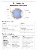
INTERNAL STRUCTURE OF THE EYE
SCLERA CORNEA:
Strong, white, inelastic connective tissue layer Front transparent portion of sclera,
that makes up outer layer of the eyeball and which is more convex than rest of the
extends to transparent cornea at the front. eyeball.
FUNCTIONS: FUNCTIONS:
• Protect internal areas of the eye. • Allow light rays to pass through to
• Helps maintain round shape of the eyeball. inner layers of the eye.
• Serves as an attachment site for muscles. • Curvature causes refraction of
incoming rays of light.
CHOROID: CILIARY BODY IRIS
Thin, dark Thickening of the frontal A round, coloured structure in
pigmented, blood part of choroid that is middle of the eye. It is at the
vessel-rich layer. made up of involuntary front of the lens & is a
muscles, known as ciliary continuation of the choroid.
FUNCTION: muscles. Ciliary body is Made up of pigments that give
Pigment absorbs attached to lens by colour to the eye, two sets of
excess light rays to suspensory ligaments. involuntary muscles, the radial
prevent internal and circular muscles.
reflection that causes FUNCTIONS
dull images. Contracts and relaxes to FUNCTION:
Blood vessels change the curvature of Regulates the amount of light
provide nutrients + the lens during eye that enters the eye, by
oxygen to the cells. accommodation. controlling the size of the pupil.
, LENS
Rubbery, elastic, clear, biconvex structure that can change shape. It is held in position by the
suspensory ligament that is attached to the ciliary body.
FUNCTION:
Changes shape to break light from far and close objects so that it forms a clear image on the
retina.
RETINA RETINA
Outer most layer that is in contact with On the retina, within the direct line of sight,
choroid. there is an area that has a lot of cone cells
Inner nervous layer is made up of two but very little rod cells, this area is called the
types of photoreceptors, namely, the cone Yellow spot. In the middle of the yellow
and rod cells. spot is the Fovea Centralis, that contains
FUNCTIONS only cone cells.
Dark pigment absorbs light rays and FUNCTIONS
prevents internal reflections. The clearest, most accurate images are
The photoreceptors observe and convert formed here.
stimuli into nerve impulses. The rod cells Nerve cells from the photoreceptors form
are stimulated in low light intensity and are synapses with sensory bipolar neurons,
responsible for black and white vision. The which are synaptically bonded to form the
cone cells are stimulated in high light optic nerve. The optic nerve leaves the
intensity and are responsible for sharp, eyeball and sends nerve impulses to the
clear, colour vision. brain.
OPTIC NERVE FUNCTIONS
Leads nerve impulses to cerebral cortex where it is interpreted and thus, we have sight.
Place where the optic nerve leaves the eye contains no rod or cone cells, is called the blind
spot. When light rays fall on this area, no nerve impulses are sent to the brain, and thus, no
images are formed.