
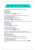
Radiology Exam Review (Appleton Lange) ARRT Questions and Answers The junction of the transverse colon and the descending colon forms the (A) hepatic flexure. (B) splenic flexure. (C) transverse flexure. (D) singmoid flexure. (Ballinger & Frank, vol 2, p 89) ✅(B) splenic flexure. All of the following statements regarding respiratory structures are true except (A) The right lung has two lobes. (B) The uppermost portion of the lung is the apex. (C) Each lung is enclosed in pleura. (D) The trachea bifurcates into mainstem bronchi. (Bontrager, pp 69 -70) ✅(A) The right lung has two lobes. When the patient is recumbent and the central ray is directed horizontally, the patient is said to be in the (A) Trendelenburg position. (B) Fowler's position. (C) decubitus position. (D) Sims position. ✅(C) decubitus position. n the AP projection of the ankle, the 1. plantar surface of the foot is vertical. 2. fibula projects more distally than the tibia. 3. calcaneus is well visualized. ✅(B) 1 and 2 only Which of the following positions would be obtained with the patient lying prone recumbent on the radiographic table, and the central ray directed horizontally to the iliac crest? (A) Ventral decubitus position (B) Dorsal decubitus position (C) Left lateral decubitus position (D) Right lateral decubitus position ✅(A) Ventral decubitus position Which of the following is (are) required for a lateral projection of the skull? 1. The IOML is parallel to the film. 2. The MSP is perpendicular to the film. 3. The central ray enters 2 in superior to the external auditory meatus (EAM). ✅(B) 1 and 3 only Which of the following should be performed to rule out subluxation or fracture of the cervical spine? (A) Oblique cervical spine, seated (B) AP cervical spine, recumbent (C) Horizontal beam lateral (D) Laterals in flexion and extension ✅C) Horizontal beam lateral Which of the following may be used to evaluate the glenohumeral joint? 1. Scapular Y projection 2. Inferosuperior axial 3. Transthoracic lateral ✅D) 1, 2, and 3 The ileocecal valve is normally located in which of the following body regions? (A) Right iliac (B) Left iliac (C) Right lumbar (D) Hypogastric ✅A) Right iliac Which of the following projections of the abdomen may be used to demonstrate air or fluid levels? 1. Dorsal decubitus 2. Lateral decubitus 3. AP Trendelenburg ✅B) 1 and 2 only The sternoclavicular joints are best demonstrated with the patient PA and (A) in a slight oblique position, affected side adjacent to the image recorder. (B) in a slight oblique position, affected side away from the image recorder. (C) erect and weight -bearing. (D) erect, with and without weights. ✅A) in a slight oblique position, affected side adjacent to the image recorder. The stomach of an asthenic patient is most likely to be located (A) high, transverse, and lateral. (B) low, transverse, and lateral. (C) high, vertical, and toward the midline. (D) low, vertical, and toward the midline. ✅D) low, vertical, and toward the midline. Deoxygenated blood from the head and thorax is returned to the heart by the (A) pulmonary artery. (B) pulmonary veins. (C) superior vena cava. (D) thoracic aorta. ✅C) superior vena cava. Which of the following statements regarding myelography is (are) correct? 1. Spinal puncture may be performed in the prone or flexed lateral position. 2. Contrast medium distribution is regulated through x -ray tube angulation. 3. The patient's neck must be in extension during Trendelenburg positions. ✅(C) 1 and 3 only Which of the following vertebral groups form(s) lordotic curve(s)? 1. Cervical 2. Thoracic 3. Lumbar ✅D) 1 and 3 only Which of the following techniques would provide a PA projection of the gastroduodenal surfaces of the barium -filled, high and transverse stomach? (A) Place the patient in a 35 to 40º RAO position. (B) Place the patient in a lateral position. (C) Angle the central ray 35 to 45º cephalad. (D) Angle the central ray 35 to 45º caudad. ✅C) Angle the central ray 35 to 45 º cephalad. Which of the following structures is (are) most likely to be demonstrated in a right lateral decubitus position of a double -contrast BE? 1. Lateral wall of the descending colon 2. Medial wall of the ascending colon 3. Lateral wall of the ascending colon (Saia, pp 180, 181) ✅C) 1 and 2 only To demonstrate esophageal varices, the patient must be examined in (A) the recumbent position. (B) the erect position. (C) the anatomic position. (D) Fowler's position. ✅A) the recumbent position. The contraction and expansion of arterial walls in accordance with forceful contraction and relaxation of the heart is called (A) hypertension. (B) elasticity. (C) pulse. (D) pressure. ✅(C) pulse. The structure(s) best demonstrated on an AP axial projection of the skull with the central ray directed 40 to 60º caudally is (are) the (A) entire foramen magnum and the jugular foramina. (B) petrous pyramids. (C) occipital bone. (D) rotundum foramina. ✅(A) entire foramen magnum and the jugular foramina. Which of the following is a radiologic procedure that functions to dilate a stenotic vessel? (A) Percutaneous nephrolithotomy (B) Percutaneous angioplasty (C) Renal arteriography (D) Surgical nephrostomy ✅(B) Percutaneous angioplasty Which of the following would be obtained with the position illustrated in Figure 2 -17? 1. Splenic flexure and descending colon 2. Hepatic flexure and ascending colon 3. Hepatic flexure and descending colon (Ballinger & Frank, vol 2, p 141) ✅(A) 1 only Which of the following is represented by the number 3 in Figure 2 -34? (A) Inferior vena cava (B) Aorta (C) Gallbladder (D) Psoas muscle (Bontrager, p 100) ✅(B) Aorta In what position was the radiograph in Figure 2 -30 made? (A) Flexion (B) Extension (C) Left bending (D) Right bending (Ballinger & Frank, vol 1, pp 398, 399) ✅A) Flexion In which of the following ways was the image seen in Figure 2 -6 obtained? (A) PA, chin extended, OML forming 37º to table (B) PA, OML and central ray (CR) perpendicular to table (C) PA, OML perpendicular to table, CR 25º caudad (D) PA, OML perpendicular to table, CR 25º cephalad (Ballinger & Frank, vol 2, pp 366 -367) ✅(C) PA, OML perpendicular to table, CR 25 º caudad Indications for a myelographic cervical puncture include 1. demonstration of the upper level of a spinal block. 2. suspected mass lesion in the upper cervical canal. 3. failure of lumbar puncture. ✅(B) 1 and 3 only Which of the following will best demonstrate the size and shape of the liver and kidneys? (A) Lateral abdomen (B) AP abdomen (C) Dorsal decubitus abdomen
Preview 4 out of 53 pages
Exam (elaborations)
Radiology Exam Review (Appleton Lange) ARRT Questions and Answers
Preview 4 out of 53 pages
The junction of the transverse colon and the descending colon forms the (A) hepatic flexure. (B) splenic flexure. (C) transverse flexure. (D) singmoid flexure. (Ballinger & Frank, vol 2, p 89) (B) splenic flexure. All of the following statements regarding respiratory structures are true except (A) The right lung has two lobes. (B) The uppermost portion of the lung is the apex. (C) Each lung is enclosed in pleura. (D) The trachea bifurcates into mainstem bronchi. (Bontrager, pp 69-70) (A) The right lung has two lobes. When the patient is recumbent and the central ray is directed horizontally, the patient is said to be in the (A) Trendelenburg position. (B) Fowler's position. (C) decubitus position. (D) Sims position. (C) decubitus position. n the AP projection of the ankle, the 1. plantar surface of the foot is vertical. 2. fibula projects more distally than the tibia. 3. calcaneus is well visualized. (B) 1 and 2 only Which of the following positions would be obtained with the patient lying prone recumbent on the radiographic table, and the central ray directed horizontally to the iliac crest? (A) Ventral decubitus position (B) Dorsal decubitus position (C) Left lateral decubitus position (D) Right lateral decubitus position (A) Ventral decubitus position Which of the following is (are) required for a lateral projection of the skull? 1. The IOML is parallel to the film. 2. The MSP is perpendicular to the film. 3. The central ray enters 2 in superior to the external auditory meatus (EAM). (B) 1 and 3 only
Content preview
Document information
- Uploaded on
- May 8, 2024
- Number of pages
- 53
- Written in
- 2023/2024
- Type
- Exam (elaborations)
- Contains
- Questions & answers
$17.49
Wrong document? Swap it for free
Within 14 days of purchase and before downloading, you can choose a different document. You can simply spend the amount again.
Written by students who passed
Immediately available after payment
Read online or as PDF

Reputation scores are based on the amount of documents a seller has sold for a fee and the reviews they have received for those documents. There are three levels: Bronze, Silver and Gold. The better the reputation, the more your can rely on the quality of the sellers work.
Available in package deal
Package deal
Comprehensive ARRT Q&A Exam Bundle

