MOLECULAR MICROBIOLOGY
BACTERIAL PHYSIOLOGY
TOPIC 1: CELL SHAPE & ARCHITECTURE
MICROBIAL SIZE & SHAPE
What pathways and structures dictate cell shape?
What structures provide cell strength?
How do bacteria respond to environment?
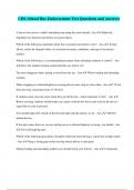
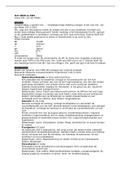
• Example E.coli:
→ SEM: via light reflection
→ TEM: light penetrating trough object
→ Rod-shaped, uniform (+ some cells longer than others = dividing cells)
→ What determines the mean size? Is there a ‘ruler’? Or a timer (like an average growth rate)?
→ Dens cytoplasm + dens boundaries (phospholipid membrane + outer membrane) + less dens periplasm
→ A flagel
• Cell shape can depend on physiological state
• Cell shape is functionally important
• Cells often adopt multicellularity, i.e. microcolonies, biofilms
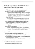
→ Eg. E.coli can bind on urotherial cells (bladder) and form intracellular cell
communities → to spread, they have to come out of the urotherial cell (lysis):
forming elongated cells (picture)
o Why elongate? To survive macrophages = a protective phenomenon
• But what regulation is behind this?
Sizes & shapes
• Spherical bacteria: + 0.5 – 2.0 µm
• Rod shaped or filamentous bacteria: + 1-100µm x + 0.25-1.0µm
• Eukaryotic cells: 10-100µm
• Viruses: 20-500 nm
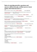
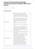
Eg. spiral Helicobacter in the stomach:
• Normally a virulent bacterium
• Stomach = pH 2 → would kill bacterium
• Solution: lives in stomach mucosa → spiral shape to stay attached to
mucosa → no motility anymore → not virulent anymore
CELL SIZE & SHAPE
• Cells without boundary = dead → many drugs target cell membrane of pathogens
• Why are they not all a sphere?
• What dictates their shape?
• Does shape matter?
• How does a cell expand in size and split during fusion?
• How do so safely without compromising cell integrity?
• What regulates all these phenomena?
• How to transport and communicate across the barrier? Eukaryotic shapes
Eukaryotes: shape is regulated from the inside: cytoskeleton, actin!
Bacteria: mostly organized from the outside: an exoskeleton
1
,LIFE IN AN UNBUFFERED ENVIRONMENT
• Osmosis = water movement from low to high concentration of
molecules via a semi-permeable membrane
• Aquaporines let water pass through the membrane
• In a confined boundary like a cell, there is pressure = ‘turgor pressure’
→ Turgor pressure in bacteria: 0.5-3 mPa (5-30 Bar)
→ In hypotonic environment: higher osmolytes outside: water flows out → plasma membrane collapse →
BUT: due to a cell wall, it retains its shape
→ (isotonic = … ; hypertonic = …)
MICROBIAL CELL ENVELOPES
Role of the cell envelope
• Rigid & protects cytoplasmic membrane
• A selective barrier that allows entry of nutrients, while reducing entry of toxic compounds, or leakage of cell
content
• Determines & supports cell size and shape
• Provides scaffold for attachment of cell appendages
• Prevents cell rupture due to osmotic challenge
• Contains functions for cell wall assembly and growth
Role of cell (cytoplasmic) membrane
• Not rigid!
• Permeability barrier
→ Even water will cross with low efficiency!
• Energy conversion; protein motive force & electron transport
→ Hydrolysable energy sources are exclusive to cytoplasm
Bacteria – Archaea – Eukarya
➔ Mycobacteria: sugar chain with fatty acids on top to make a pseudomembrane → difficult to combat
BACTERIAL CELL ENVELOPES
2
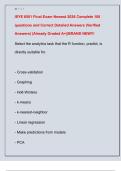
,GRAM-POSITIVE CELL ENVELOPES
• Gram positive or monoderm bacteria
• Thick 20-80 nm
• Peptidoglycan layer, + 50% of cell envelop
• No lipids
• Secondary cell wall polymers: teichoic acid and/or teichuronic acid
→ Covalently linked to PG / anchored in the cytoplasmic
membrane
• PG-bound surface proteins: eg. pili, cell recognition proteins…
→ Eg. if they live on plant material: polymers to degrade lignine/cellulose/… (= large polymers) = eg.
cellulases → why anchored? Otherwise other bacteria could make advantage of it
• Good permeability
TEICHOIC ACIDS
• Covalently attached to PG (phosphodiester to MurNAc)
• Account for as much as 60% of total cell wall mass
• ManNac(β1→4)GlcNac disaccharide with one to three glycerol phosphates attached to the C4 hdyroxyl of
the ManNac residue (the ‘linkage unit’) followed by a much longer chain of glycerol- or ribitol phosphate
repeats (main chain)
• So, a linkage to sugar units and a polymer of eiter glycerol or ribitol P → so quite negatively charged
• Virulence associated, i.e. adherence and biofilm formation (eg. MRSA)
• You don’t have to be able to draw it
LIPOTECHOIC ACIDS
• 4 types, lipid anchored techoic acids found
→ Eg. polyglycerolphosphate (type I LTA): best-characterized LTA
o In phylum Firmicutes: eg. Staphylococcus aureus, Bacillus subtilus, Listeria monocytogenes
• Plays important role for bacterial growth and physiology
• Contributes to membrane homeostasis and virulence
→ So this makes LTA a target for vaccines and novel antimicrobial drugs
• LTA modification can protect against antimicrobial peptides
GRAM-NEGATIVE CELL ENVELOPES
• Gram negative or diderm bacteria
• Thin 8-11 nm
• PG monolayer
• Second lipid bilayer: outer membrane
→ Asymmetric with inner phospholipid layer + outer lipopolysaccharide layer (LPS)
→ Essential
• Periplasm < periplasmic proteins (up to 10% of proteome)
→ Oxidizing environment
→ No hydrolysable energy source there (like NAD etc. = inside cytoplasm)
→ So no trivial functions there
• Lipoproteins: inner & outer membrane
→ Specific sorting system → LOL pathway
• Outer membrane proteins → beta barrels
• Dedicated secretion and import systems (see Chap.3) Because molecules have to pass 2 membranes
• Less permeable → less susceptible to antibiotics
3
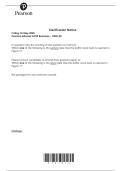
, OUTER MEMBRANE
• An additional permeability barrier
• Multitude secretion/import system (C3)
• Lot of proteins in outer membrane: mostly porines
→ Porin-based passive passage: + 500 Da molecules
• No ion gradients
• LPS remodeled in function of antibiotic pressure
→ O-antigen variation to escape immune response
→ LPS anchored on membrane via lipid A
o Lipid A = endotoxin → recognized by toll like
receptor 4 (innate IMS)
• Intrinsic resistance to antibacterials
→ Many AB’s are lipophilic → but LPS is negatively
charged so often AB’s cannot pass through it
→ Polymyxin, bile salts
→ Antibacterial peptides: bind membrane and makes pores → bacterium can modify lipids/phosphates so
that they are less attacked
• Mg and Ca2+ in outer membrane
2+
→ Many stressors remove these (eg. during fagocytosis)
o Sometimes, the bacterium modifies itself so that it does not need Mg for example anymore
OUTER MEMBRANE BIOSYNTHESIS
• Pathways are needed to get the proteins/molecules/… in
the membranes
• LPS synthesis at the inner membrane via Lpx pathway
→ Eg. LPS flipase: transporting LPS from IM to OM
• Transport to outer membrane via Lpt pathway
• Pathway is essential
• Outer membrane phospholipids ?
→ Not known how they get there
→ Has to be very coordinated: one fault = symmetry gone = membrane broken
• No outer membrane = cell dies
→ But why? All functions are in inner membrane, so? Not known
→ Good drug target
OUTER MEMBRANE STABILITY
• PG and outer membrane to give cell stability → °turgor pressure
• Loss of OM stability = cell lysis under hypotonic conditions
• 2 component system
→ Sensor kinase (stress senor) + response regulator
→ Transcription regulation
• Can sense destabilizing things in the OM: eg. Mg2+ taken away → but cell can take away the need for Mg2+
→ Attack by antimicrobial peptides (IMS) → bacterium itself will
take away Mg2+’s → antimicrobial peptides can no longer
detect Mg2+
o How: via LPS remodeling enzymes: replacing the LPS by
phospholipids
→ Plus: overproduction of SlyB = an OM guard protein, giving
rigidity to membrane → turgor pressure
4
BACTERIAL PHYSIOLOGY
TOPIC 1: CELL SHAPE & ARCHITECTURE
MICROBIAL SIZE & SHAPE
What pathways and structures dictate cell shape?
What structures provide cell strength?
How do bacteria respond to environment?
• Example E.coli:
→ SEM: via light reflection
→ TEM: light penetrating trough object
→ Rod-shaped, uniform (+ some cells longer than others = dividing cells)
→ What determines the mean size? Is there a ‘ruler’? Or a timer (like an average growth rate)?
→ Dens cytoplasm + dens boundaries (phospholipid membrane + outer membrane) + less dens periplasm
→ A flagel
• Cell shape can depend on physiological state
• Cell shape is functionally important
• Cells often adopt multicellularity, i.e. microcolonies, biofilms
→ Eg. E.coli can bind on urotherial cells (bladder) and form intracellular cell
communities → to spread, they have to come out of the urotherial cell (lysis):
forming elongated cells (picture)
o Why elongate? To survive macrophages = a protective phenomenon
• But what regulation is behind this?
Sizes & shapes
• Spherical bacteria: + 0.5 – 2.0 µm
• Rod shaped or filamentous bacteria: + 1-100µm x + 0.25-1.0µm
• Eukaryotic cells: 10-100µm
• Viruses: 20-500 nm
Eg. spiral Helicobacter in the stomach:
• Normally a virulent bacterium
• Stomach = pH 2 → would kill bacterium
• Solution: lives in stomach mucosa → spiral shape to stay attached to
mucosa → no motility anymore → not virulent anymore
CELL SIZE & SHAPE
• Cells without boundary = dead → many drugs target cell membrane of pathogens
• Why are they not all a sphere?
• What dictates their shape?
• Does shape matter?
• How does a cell expand in size and split during fusion?
• How do so safely without compromising cell integrity?
• What regulates all these phenomena?
• How to transport and communicate across the barrier? Eukaryotic shapes
Eukaryotes: shape is regulated from the inside: cytoskeleton, actin!
Bacteria: mostly organized from the outside: an exoskeleton
1
,LIFE IN AN UNBUFFERED ENVIRONMENT
• Osmosis = water movement from low to high concentration of
molecules via a semi-permeable membrane
• Aquaporines let water pass through the membrane
• In a confined boundary like a cell, there is pressure = ‘turgor pressure’
→ Turgor pressure in bacteria: 0.5-3 mPa (5-30 Bar)
→ In hypotonic environment: higher osmolytes outside: water flows out → plasma membrane collapse →
BUT: due to a cell wall, it retains its shape
→ (isotonic = … ; hypertonic = …)
MICROBIAL CELL ENVELOPES
Role of the cell envelope
• Rigid & protects cytoplasmic membrane
• A selective barrier that allows entry of nutrients, while reducing entry of toxic compounds, or leakage of cell
content
• Determines & supports cell size and shape
• Provides scaffold for attachment of cell appendages
• Prevents cell rupture due to osmotic challenge
• Contains functions for cell wall assembly and growth
Role of cell (cytoplasmic) membrane
• Not rigid!
• Permeability barrier
→ Even water will cross with low efficiency!
• Energy conversion; protein motive force & electron transport
→ Hydrolysable energy sources are exclusive to cytoplasm
Bacteria – Archaea – Eukarya
➔ Mycobacteria: sugar chain with fatty acids on top to make a pseudomembrane → difficult to combat
BACTERIAL CELL ENVELOPES
2
,GRAM-POSITIVE CELL ENVELOPES
• Gram positive or monoderm bacteria
• Thick 20-80 nm
• Peptidoglycan layer, + 50% of cell envelop
• No lipids
• Secondary cell wall polymers: teichoic acid and/or teichuronic acid
→ Covalently linked to PG / anchored in the cytoplasmic
membrane
• PG-bound surface proteins: eg. pili, cell recognition proteins…
→ Eg. if they live on plant material: polymers to degrade lignine/cellulose/… (= large polymers) = eg.
cellulases → why anchored? Otherwise other bacteria could make advantage of it
• Good permeability
TEICHOIC ACIDS
• Covalently attached to PG (phosphodiester to MurNAc)
• Account for as much as 60% of total cell wall mass
• ManNac(β1→4)GlcNac disaccharide with one to three glycerol phosphates attached to the C4 hdyroxyl of
the ManNac residue (the ‘linkage unit’) followed by a much longer chain of glycerol- or ribitol phosphate
repeats (main chain)
• So, a linkage to sugar units and a polymer of eiter glycerol or ribitol P → so quite negatively charged
• Virulence associated, i.e. adherence and biofilm formation (eg. MRSA)
• You don’t have to be able to draw it
LIPOTECHOIC ACIDS
• 4 types, lipid anchored techoic acids found
→ Eg. polyglycerolphosphate (type I LTA): best-characterized LTA
o In phylum Firmicutes: eg. Staphylococcus aureus, Bacillus subtilus, Listeria monocytogenes
• Plays important role for bacterial growth and physiology
• Contributes to membrane homeostasis and virulence
→ So this makes LTA a target for vaccines and novel antimicrobial drugs
• LTA modification can protect against antimicrobial peptides
GRAM-NEGATIVE CELL ENVELOPES
• Gram negative or diderm bacteria
• Thin 8-11 nm
• PG monolayer
• Second lipid bilayer: outer membrane
→ Asymmetric with inner phospholipid layer + outer lipopolysaccharide layer (LPS)
→ Essential
• Periplasm < periplasmic proteins (up to 10% of proteome)
→ Oxidizing environment
→ No hydrolysable energy source there (like NAD etc. = inside cytoplasm)
→ So no trivial functions there
• Lipoproteins: inner & outer membrane
→ Specific sorting system → LOL pathway
• Outer membrane proteins → beta barrels
• Dedicated secretion and import systems (see Chap.3) Because molecules have to pass 2 membranes
• Less permeable → less susceptible to antibiotics
3
, OUTER MEMBRANE
• An additional permeability barrier
• Multitude secretion/import system (C3)
• Lot of proteins in outer membrane: mostly porines
→ Porin-based passive passage: + 500 Da molecules
• No ion gradients
• LPS remodeled in function of antibiotic pressure
→ O-antigen variation to escape immune response
→ LPS anchored on membrane via lipid A
o Lipid A = endotoxin → recognized by toll like
receptor 4 (innate IMS)
• Intrinsic resistance to antibacterials
→ Many AB’s are lipophilic → but LPS is negatively
charged so often AB’s cannot pass through it
→ Polymyxin, bile salts
→ Antibacterial peptides: bind membrane and makes pores → bacterium can modify lipids/phosphates so
that they are less attacked
• Mg and Ca2+ in outer membrane
2+
→ Many stressors remove these (eg. during fagocytosis)
o Sometimes, the bacterium modifies itself so that it does not need Mg for example anymore
OUTER MEMBRANE BIOSYNTHESIS
• Pathways are needed to get the proteins/molecules/… in
the membranes
• LPS synthesis at the inner membrane via Lpx pathway
→ Eg. LPS flipase: transporting LPS from IM to OM
• Transport to outer membrane via Lpt pathway
• Pathway is essential
• Outer membrane phospholipids ?
→ Not known how they get there
→ Has to be very coordinated: one fault = symmetry gone = membrane broken
• No outer membrane = cell dies
→ But why? All functions are in inner membrane, so? Not known
→ Good drug target
OUTER MEMBRANE STABILITY
• PG and outer membrane to give cell stability → °turgor pressure
• Loss of OM stability = cell lysis under hypotonic conditions
• 2 component system
→ Sensor kinase (stress senor) + response regulator
→ Transcription regulation
• Can sense destabilizing things in the OM: eg. Mg2+ taken away → but cell can take away the need for Mg2+
→ Attack by antimicrobial peptides (IMS) → bacterium itself will
take away Mg2+’s → antimicrobial peptides can no longer
detect Mg2+
o How: via LPS remodeling enzymes: replacing the LPS by
phospholipids
→ Plus: overproduction of SlyB = an OM guard protein, giving
rigidity to membrane → turgor pressure
4