

Chapter 5: The Integumentary System
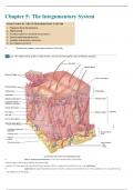
Chapter 5: The Integumentary System 1. Describe the anatomy and major functions of the skin. • skin or cutaneous membrane-covers the external surface of the body. -largest organ of the body(in surface area and weight) -consists of 2 main parts: the superficial thinner portion which is composed of epithelial tissue is the epidermis. • epidermis-the superficial thinner portion which is composed of epithelial tissue is the epidermis. It is avascular (if you cut epidermis=no bleeding) • dermis-deeper, thicker connective tissue portion is the dermis. It is vascular (if you cut dermis=bleeding) • subcutaneous (SQ) layer or hypodermis- deep to the dermis, but not part of the skin, ist he subcutaneous (subQ) layer. -Also called the hypodermis, this layer consists of areolar and adipose tissues. -Fibers that extend from the dermis anchor the skin to the subcutaneous layer, which in turn attaches to underlying fascia, the connective tissue around muscles and bones. -The subcutaneous layer serves as a storage depot for fat and contains large blood vessels that supply the skin. • Epidermis- composed of keratinized stratified squamous epithelium. -contains 4 principal types of cells: keratinocytes, melanocytes, Langerhans cells, and Merkel cells • Keratinocyte-90% of epidermal cells are keratinocytes. -arranged in 4 or 5 layers and produce the protein keratin -also produce lamellar granules, which release a water-repellent sealant that decreases water entry and loss and inhibits the entry of foreign materials. • Keratin-a tough, fibrous protein that helps protect the skin and underlying tissues from abrasions, heat, microbes, and chemicals. • Melanocyte-8% of epidermal cells are melanocytes -develop from the ectoderm of a developing embryo and produce the pigement melanin. -Their long, slender projections extend between the keratinocytes and transfer melanin granules to them. • Melanin-is a yellow-red or brown-black pigment that contributes to skin color and absorbs damaging ultraviolet (UV) light. -once inside keratinocytes, the melanin granules cluster to form a protective veil over the nucleus, on the side toward the skin surface. In this way, they shield the nuclear DNA from damage by UV light. -Although their melanin granules effectively protect keratinocytes, melanocytes themselves are particularly susceptible to damage by UV light. • Langerhans cell-also called epidermal dendritic cells, arise from red bone marrow and migrate to the epidermis where they constitute a small fraction of the epidermal cells -they participate in immune responses mounted against microbes that invade the skin, and are easily damaged by UV light. -their role in the immune response is to help other cells of the immune system recognize an invading microbe and destroy it. • Merkel cell-least numerous of the epidermal cells -located in the deepest layer of the epidermis, where they contact the flattened process of a sensory neuron (nerve cell), a structure called a Merkle (tactile) disc. -Merkel cells and their associated Merkel discs detect tough sensations. • layers or strata-in most regions of the body the epidermis has four strata or layers- stratum basal, stratum spinosum, stratum granulosum, and a thin stratum corneum. This is called thin skin. -Where exposure to friction is greatest, such as in fingertips, palms, and soles, the epidermis has 5 layers: stratum basal, stratum spinosum, stratum granulosum, stratum lucidum, and a thick stratum corneum. This is called thick skin. • stratum basale or stratum germinativum- deepest layer of epidermis -composed of a single row of cuboidal or columnar keratinocytes. -Some cells in this layer are stem cells that undergo cell division to continually produce new keratinocytes. -Nuclei of keratinocytes in the stratum basale are large, and their cytoplasm contains many ribosomes, a small Golgi complex, a few mitochondria, and some rough endoplasmic reticulum. -The cytoskeleton within keratinocytes of the stratum basale includes scattered intermediate filaments, called keratin intermediate filaments (tonofilaments). They form the tough protein keratin in its more superficial epidermal layers. -keratin protects the deeper layers from injury. -Keratin intermediate filaments attach to desmosomes, which bind cells of the stratum basale to each other and to the cells of the adjacent stratum spinosum, and to hemidesmosomes, which bind keratinocytes to the basement membrane positioned between the epidermis and the dermis. -Melanocytes and Merkel cells with their associated Merkel discs are scattered among the keratinocytes of the basal layer. -statum basale is also known as the statum germinativum to indicate its role in forming new cells. • stratum spinosum -superficial to the statum basale -mainly consists of numerous keratinocytes arranged in 8-10 layers. -Cells in the more superficial (top) layers become somewhat flattened. -Keratinocytes in the stratum spinosum are produced by the stem cells in the basal layer. They have the same organelles as cells of the stratum basale, and some retain their ability to divide. -Keratinocytes of this layer produce coarser bundles of keratin in intermediate filaments than those of the basal layer -round and larger in living tissue, but they shrink and pull apart when prepared for microscopic exams. They appear to be covered with thornlike spines -At each spinelike projection, bundles of keratin intermediate filaments insert into desmosomes, which tightly join the cells to one another. This arrangement provides strength and flexibility to skin -Langerhans cells and projections of melanocytes are also present in the stratum spinosum. • stratum granulosum-middle of epidermis -consists of 3-5 layers of flattened keratinocytes that are undergoing apoptosis (an orderly, genetically programmed cell death in which the nucleus fragments before the cells die). The nuclei and other organelles of these cells begin to degenerate as they move farther from their source of nutrition (the dermal blood vessels) -even though keratin intermediate filaments are no longer being produced by these cells, they become more apparent because the organelles in the cell are regressing. -As their nuclei break down during aptopsis, the keratinocytes of the stratum granulosum can no longer carry on vital metabolic reactions, and they die. -Thus, the stratum granulosum marks the transition between the deeper, metabolically active strata, and the dead cells of the more superficial strata. • stratum lucidum-present only in the thick skin of areas (fingertips, palms, and soles) -consists of 4-6 layers of flattened clear, dead keratinocytes that contain large amounts of keratin and thickened plasma membranes. -Probably provides an additional level of toughness in this region of thick skin • stratum corneum- consists on avg of 25-30 layers of flattened dead keratinocytes, but can range in thickness from a few cells in thin skin to 50 or more cell layers in thick skin. -cells are extremely thin, flat, plasma membrane-enclosed packages of keratin that no longer contain a nucleus or any internal organelles. They are the final product of the differentiation process of the keratinocytes -Cells within each layer overlap one another like scales on a snake -Neighboring layers of cells also form strong connections with one another. The plasma membranes of adjacent cells are arranged in complex, wavy folds that fit together like pieces of a jigsaw puzzle to hold the layers together. -cells are continuously shed and replaced by cells from the deeper strata. -Its multiple layers of dead cells help the stratum corneum to protect deeper layers from injury and microbial invasion. -constant exposure of skin to friction stimulates increased cell production and keratin production that results in the formation of a callus, an abnormal thickening of the stratum corneum. • Keratinization-Newly formed cells in the stratum basal are slowly pushed to the surface. As the cells move from one epidermal layer to the next, they accumulate more and more keratin, a process called keratinization. Then they undergo apoptosis. -eventually the keratinized cells slough off and are replaced by underlying cells that in turn become keratinized. -the whole process takes about 4-6 weeks in an avg epidermis of 0.1mm thickness. -cells in the stratum basale layer are closest to blood vessels and receive most of the nutrients and oxygen. They are the most active metabolically and continuously undergo cell division to produce new keratinocytes. -The rate of cell division in the stratum basale increases when the otuer layers of the epidermis are stripped away, as occurs in abrasions and burns. • Dermis-second deeper part of skin is composed of dense irregular connective tissue containing collagen and elastic fibres. -woven network of fibres has great tensile strength (resist pulling or stretching forces). -also has the ability to stretch and recoil easily -Much thicker than epidermis -Leather is the dried and treated dermis of other animals. -Few cells present in dermis include: predominantly fibroblasts, with some macrophages, and a few adipocytes near its boundary with the subcutaneous layer. -blood vessels, nerves, glands, and hair follicles (epithelial invaginations of the epidermis) are embedded in the dermal layer. -dermis is essential to survival of epidermis, and these adjacent layers form many important structural and functional relations. -can be divided into a thin superficial papillary region and a thick deeper reticular region. • papillary region-makes up about 1/5 of the thickness of the total layer. -Consists of thin collagen and fine elastic fibres -its surface area is greatly increased by dermal papillae • dermal papilla (plural is papillae)-small, nipple-shaped structures that project into the undersurface of the epidermis. -all dermal papilla contain capillary loops (blood vessels) Some also contain tactile receptors. • corpuscle of touch or Meissner corpuscle-tactile receptors. Nerve endings that are sensitive to touch • free nerve ending-other dermal papillae also contain free nerve endings, dendrites that lack any apparent structural specialization. -different free nerve endings initiate signals that give rise to sensations of warmth, coolness, pain, tickling, and itching. • reticular region-attached to the subcutaneous layer, consists of bundles of thick collagen fibres, scattered fibroblasts, and various wandering cells (such as macrophages). -some adipose cells can be present in the deepest part of the layer, along with some coarse elastic fibres. -collagen fibres are arranged in a netlike manner and have more regular arrangement than those in the papillary region. The more regular orientation of the large collagen fibres helps the skin resist stretching. -Blood vessels, nerves, hair follicles, sebaceous glands (oil glands), and sudoriferous (sweat) glands occupy the spaces between fibres. • epidermal ridge-surfaces of the palms, fingers, soles and toes have a series of ridges and grooves. They appear as straight lines, or as a pattern of loops and whorls. -These epidermal ridges are produced during the 3rd month of fetal development as downward projections of the epidermis into the dermis between the dermal papillae of the papillary region. -create a strong bond between the epidermis and dermis in a region of high mechanical stress. -increases the surface area of the epidermis and thus increase the grip of the hand or foot by increasing friction. -greatly increases surface area, which increases the number of Messiner corpuscles and thus increasing tactile sensitivity. -ducts of sweat glands open on the tops of epidermal ridges as sweat pores, the sweat and ridges form fingerprints on touching a smooth object. -in part genetically determined. Unique for each individual. Identical twins have different patterns. -basis for identification 2. Explain the basis of different skin colours. • structural basis of skin colour-melanin, hemoglobin, and carotene are 3 piggments that impart a wide variety of colors to skin • melanin-causes the skin’s color to vary from pale yellow to reddish-brown to black. The difference between the two forms of melanin, pheomelanin(yellow to red) and eumelanin(brown to black) is most apparent in the hair. -melanocytes, the melanin producing cells, are most plentiful in the epidermis of the penis, nipples of the breasts, area just around the nipples (areolae), face, and limbs. Also present in mucous membranes. -because the number of melanocytes is about the same in all people, the difference in skin color are due mainly to the amount of piegment the melanocytes produce and transfer to keratinocytes. -can accumulate in patches called freckles. And age spots. • Carotene-a yellow-orange pigment that gives egg yolks and carrots their color -This precursor of vitamin A, which is used to synthesize pigments needed for vision, is stored in the stratum corneum and fatty areas of the dermis and subcutaneous layer in response to excessive dietary intake. -so much carotene may be deposited in the skin after eating large amounts of carotene-rich foods and the skin actually turns orange. • Albinism- inherited inability of an individual to produce melanin. Most have melanocytes that are unable to synthesize tyrosinase. Melanin is missing fromt heir hair, eyes, and skin. This results in problem with vision, and a tendency of the skin to burn easily on overexposure to sunlight. 3. Compare the anatomy, distribution and physiology of the epidermal derivatives listed below. • hair • sebaceous glands or oil glands • sudoriferous glands or sweat glands • ceruminous glands • nails • Hair- hair on head guards scalp from injury and the sun’s rays. Also decreases heat loss from the scalp. -eyebrows and eyelashes protect eyes form foreign particles. -Touch receptors (hair root plexuses) associated with hair follicles are activated whenever a hair is moved even slightly. Hairs also function in sensing light touch. Anatomy of hair: -each hair is composed of columns of dead, keratinized epidermal cells bonded together by extracellular proteins. -shaft-superficial portion of the hair- protects above the surface of the skin. -root- portion of the hair deep to the shaft that penetrates into the dermis, and sometimes into the subcutaneous layer. -shaft and root consist of 3 concentric layers of cells: medulla, cortex, and cuticle of the hair. -inner medulla- may be lacking in thinner hair, is composed of 2 or 3 rows of irregularly shaped cells that contain large amounts of pigment granules in dark hair, small amounts of pigment granules in gray hair, and a lack of pigment granules and the presence of air bubbles in white hair. -middle cortex- forms the major part of the shaft and consists of elongated cells. -cuticle of the hair- the outermost layer- consists of a single layer of thin, flat cells that are most heavily keratinized. Arranged like shingles on the side of a house with their free edges prointing towards the end of the hair. -surrounding the root of the hair is the hair follicle-made up of an external root sheath and an internal root sheath, together referred to as epithelial root sheath. -external root sheath-downward continuation of the epidermis -internal root sheath- produced by the matrix and forms a cellular tubular sheath of epithelium between the external root sheath and the hair -the dense dermis surrounding the hair follicle is called the dermal root sheath. -base of each hair follicle and its surrounding root sheath is an onion-shaped structure, the bulb. It houses a nipple-shaped indentation, the papilla of the hair which contains areolar connective tissue and many blood vessels that nourish the growing hair follicle. The bubl also contains a germinal layer of cells called the hair matrix. -the hair matrix cells arise from the stratum basale, the site of cell division. Hence, hair matrix cells are responsible for the growth of existing hairs, and they produce new hairs when old ones shed. This replacement process occurs within the same follicle. Hair matrix cells also give rise to the cells of the internal root sheath. -Sebaceous (oil) glands and a bundle of smooth muscle cells are associated with hairs. The smooth muscle is the arrector pili. It extends from the superficial dermis of the skin to the dermal root sheath around the side of the hair follicle. -normally, hair emerges at an angle to the surface of the skin, but when its cold or under emotional stress, autonomic nerve endings stimulate the arrector pili muscles to contract, which pulls the hair shafts perpendicular to the skin (goose bumps). Kin around the shaft forms slight elevations. -Surrounding each hair follicle are dendrites of neurons that form a hair root plexux- which is sensitive to touch. It generates nerve impulses if the hair shafts are moved. Hair Growth -growth stage (2-6 years)- cells of the hair matrix divide. New cells are added to the base of the hair root, and existing cells of the hair root are pushed upwards and the hair grows longer. When the cells of the hair are being pushed upward, they become karitinized and die. -Regression stage(2-3 weeks)- cells of the hair matrix stop dividing, the hair follicle atrophies (shrinks), and the hair stops crowing. -resting stage (3 months) - following regression stage. After resting stage, a new growth cycle begins. -old hair root falls out or is pushed out of the hair follicle, and new hair begins to grow. -terminal hairs- prior to birth, the lanugo (very fine, nonpigmented downy hairs that are produced by follicles around 5th month of development) of the eyebrows, eyelashes, and scalp are shed and replaced by long, coars, heavily pigmented hairs called terminal hairs. -Vellus hairs- the lanugo of the rest of the body are replaced by vellus hairs- short fine pale hairs that are barely visible to the naked eye (peach fuzz) -Hair color is due primarily to the amount and type of melanin in its keratinized cells. Melanin is synthesized by melanocytes scattered in the matrix of the bulb and passes into cells of the cortex and medulla of the hair -Dark colored hair contains mostly eumelanin (brown to black) -blond and red hair contains varients of pheomelanin (yellow to red) -hair coloring is a process that adds or removes pigment. Temporary hair dyes coat the surface of a hair shaft and usually wash out within 2 or 3 shampoos. Semipermanent dies penetrate the hair shaft moderately and do fade and wash out after about 5-10 shampoos. Permanent hair dyes penetrate deeply into hair shaft and don’t wash out but are eventually lost as the hair grows out.
Document information
- Uploaded on
- September 19, 2023
- Number of pages
- 14
- Written in
- 2023/2024
- Type
- Exam (elaborations)
- Contains
- Questions & answers


