

MLS ASCP Hematology|2023 LATEST UPDATE|GUARANTEED SUCCESS
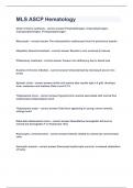
Order of heme synthesis Porphobilinogen, Uroporphyrinogen, Coproporphyrinogen, Protoporphyrinogen Macrocytic The characteristic erythrocyte found in pernicious anemia Idiopathic Hemochromatosis Results in iron overload in tissues Phlebotomy treatment Causes iron deficiency due to blood loss Anemia of chronic infection Characterized by decreased serum iron levels Aplastic crisis sickle cell anemia labs results hgb is 8 g/dL develops fever, weakness and malaise. Retic count 0.1% Thalassemia minor Hypochromic anemia associated with normal free erythrocyte protoporphyrin level Thalassemia major Fatal form appearing in young, severe anemia, enlarge heart Delta-beta thalassemia minor Quantitative hemoglobin A2 level is normal but hemoglobin F is 5%(normal 2%) Normocytic, normochromic Anemia related to uremia has normal sized cells Hemolytic anemia Decreased erythrocyte survival; increased catabolism of heme G-6-PD deficiency An enzyme deficiency associated with a moderate to severe hemolytic anemia after the patient is exposed to certain drugs Positive DAT Seen in acquired hemolytic anemias of the autoimmune variety Macrocytosis Characteristic morphologic feature in folic acid deficiency Megaloblastic anemias is caused by impaired DNA synthesis; lab results include low platelet and neutrophil counts Erythroleukemia Characteristic morphologic feature giant, vacuolated, multinucleated erythroid precursors DiGuglielmo syndrome Feature megaloblastoid erythropoiesis M:E ratio Low M:E ratio is seen in erythroleukemia; normal in polycythemia vera Polycythemia vera Decreased or absent bone marrow iron stores; elevated Hgb, Hct, and RBCs Plasmodium vivax Smear reveals Schuffner dots, parasites with irregular "spread-out" trophozoites, golden-brown pigment beta-thalassemia Characteristic elevated A2 hemoglobin alpha-thalassemia Characteristic decreased A2 hemoglobin Hemoglobin H disease Occurs when 3 of 4 alpha genes are deleted E trait Shows 70% Hgb A and 30% of a hemoglobin with the mobility Hgb A2 Fibroblast The peripheral blood monocyte is the intermediate stage in the formation of fibroblasts Basophils Involved in immediate hypersensitivity reactions M4 FAB classification of myelomonocytic leukemia Myelofibrosis with myeloid metaplasia Dwarf or micro megakaryocytes found in blood smear Auer bodies Predominately found in acute myelogenous leukemia Philadelphia chromosome Absence of Philadelphia chromosome in granulocytic leukemia suggests rapid progression of disease Granulocytic hyperplasia A hypercellular marrow with an M:E ratio of 6:1 is common Acute Myelomonocytic Leukemia Most predominant for of secondary hematologic malignancy in patients with multiple myeloma
Document information
- Uploaded on
- June 15, 2023
- Number of pages
- 5
- Written in
- 2022/2023
- Type
- Exam (elaborations)
- Contains
- Questions & answers


