
Osteologie van het onderste
lidmaat
Femur
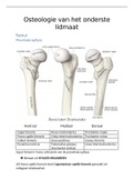
Proximale epifyse
Ventraal Mediaal Dorsaal
Caput femoris Fossa trochanterica Trochanter major
Fovea capitis femoris Crista intertrochanterica Trochanter minor
Collum femoris Linea pectinea Corpus femoris
Foramina nutricia Tuberositas glutaea Linea intertrochanterica
(trochanter tertius)
Caput femoris= facies articularis van de proximale epifyse
Bestaat uit HYALIEN KRAAKBEEN
Uit fovea capitis femoris komt Ligamentum capitis femoris gemaakt uit
collageen bindweefsel.
,Diafyse of corpus
Duidelijke richel in het bot: Linea aspera, bestaat uit labium mediaal en labium
lateraal
Naar proximaal loopt uit in Linea pectinea en Tuberositas glutaea
Naar distaal loopt uit in Linea supracondylaris lateraal en linea
supracondylaris mediaal
Tussen deze 2 ligt een afgeplatte, driehoekige opp. de Facies popliteus
Distale epifyse
Ventraal Dorsaal
Condylus lateralis Epicondylus lateralis Linea intercondylaris
Condylus medialis Epicondylus medialis Fossa intercondylaris
Facies patellaris Tuberculum Sulcus popliteus
adductorium
, Collo-diafysaire hoek= hoek tussen
nek en hoofd van femur
Normaal: ± 125°
Coxa vara: <120°
Coxa valga: >135°
Antetorsie van de femur:
proximale epifyse staat in een hoek
van ± 12° met de distale epifyse
Patella
=os sesamoïdeum (sesambot): bot omwikkeld door bindweefsel
Facies anterior Facies articularis
Apex patellae Basis patellae
Facies articularis in zijn proximale ¾ bedekt met kraakbeen
Kraakbeen in bepaalde structuur dikker aan laterale kant dan mediale
WIBERG Classificatie: classificatie dikte van kraakbeen
Type 1 Type 2 Type 3
M=L (meer problemen) L>M (norm) L>>M (meer problemen)
lidmaat
Femur
Proximale epifyse
Ventraal Mediaal Dorsaal
Caput femoris Fossa trochanterica Trochanter major
Fovea capitis femoris Crista intertrochanterica Trochanter minor
Collum femoris Linea pectinea Corpus femoris
Foramina nutricia Tuberositas glutaea Linea intertrochanterica
(trochanter tertius)
Caput femoris= facies articularis van de proximale epifyse
Bestaat uit HYALIEN KRAAKBEEN
Uit fovea capitis femoris komt Ligamentum capitis femoris gemaakt uit
collageen bindweefsel.
,Diafyse of corpus
Duidelijke richel in het bot: Linea aspera, bestaat uit labium mediaal en labium
lateraal
Naar proximaal loopt uit in Linea pectinea en Tuberositas glutaea
Naar distaal loopt uit in Linea supracondylaris lateraal en linea
supracondylaris mediaal
Tussen deze 2 ligt een afgeplatte, driehoekige opp. de Facies popliteus
Distale epifyse
Ventraal Dorsaal
Condylus lateralis Epicondylus lateralis Linea intercondylaris
Condylus medialis Epicondylus medialis Fossa intercondylaris
Facies patellaris Tuberculum Sulcus popliteus
adductorium
, Collo-diafysaire hoek= hoek tussen
nek en hoofd van femur
Normaal: ± 125°
Coxa vara: <120°
Coxa valga: >135°
Antetorsie van de femur:
proximale epifyse staat in een hoek
van ± 12° met de distale epifyse
Patella
=os sesamoïdeum (sesambot): bot omwikkeld door bindweefsel
Facies anterior Facies articularis
Apex patellae Basis patellae
Facies articularis in zijn proximale ¾ bedekt met kraakbeen
Kraakbeen in bepaalde structuur dikker aan laterale kant dan mediale
WIBERG Classificatie: classificatie dikte van kraakbeen
Type 1 Type 2 Type 3
M=L (meer problemen) L>M (norm) L>>M (meer problemen)


