

Mitochondria & peroxisomes
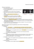
Structure of a mitochondrion
● Have outer and inner membrane
● The inner membrane space.
● The inner membrane forms the cristae. These are for increasing
S.A respiration
● Most enzymes work in the matrix of the inner membrane.
● 80% more by mass than protein & are quite plastic to the rigid
belief.
● Mitochondria often associate with microtubules [part of
cytoskeleton]
● The mitochondria move along the microtubules which help
distribution of the organelles, especially for when mitosis occurs.
Experiments of a Rod Spherule Mitochondria; 3D electron tomograms
● Rod spherule: A specialised neuron projection which is the site of synaptic transmission produced by
retinal rod cells. Rod spherules are small round enlargements of the axon (3-5 micrometers diameter) or
even extensions of the cell body.
● Cut the mitochondria in very fine specimens & used electron tomograms. Then, put the 2D pictures
together to form a 3D shape of the mitochondria.
Mitochondrial morphology changes during.[Morphology; study of the forms of things particularly
form/shape/structure]
● Apoptosis
○ When cell is stressed it may undergo DNA damage/ viral infection/ growth factor deprivation
○ If the cell is going under apoptosis, it will send signals through the mitochondria known as intrinsic
pathway.
○ When the signal is sent, proapoptotic proteins are sent to form oligomers in the outer membranes
of mitochondria.
○ The oligomers, release cytochrome C from outer to inner mitochondrial membrane.
○ The cytochrome C drives apoptosis.
2+
● Ca transfer
○ Cell stimulation with histamine, leading the activation of GQ pathway. The GQ is made from α, β,
γ subunits to make a protein. If it’s GQ, then it’s a different version of the alpha subunit.
○ The GQ pathway enables Ca2+ ions to be signalled.
● Cell cycle
○ For the S-phase, all the organelles need to be duplicated & the mitochondria is needed for ATP
production.
○ Mitosis & cytokinesis, ATP os needed.
● Nutrient starvation
○ ATP production is key for catalytic processes like glycogenolysis.
Structure of a mitochondrion
● Have outer and inner membrane
● The inner membrane space.
● The inner membrane forms the cristae. These are for increasing
S.A respiration
● Most enzymes work in the matrix of the inner membrane.
● 80% more by mass than protein & are quite plastic to the rigid
belief.
● Mitochondria often associate with microtubules [part of
cytoskeleton]
● The mitochondria move along the microtubules which help
distribution of the organelles, especially for when mitosis occurs.
Experiments of a Rod Spherule Mitochondria; 3D electron tomograms
● Rod spherule: A specialised neuron projection which is the site of synaptic transmission produced by
retinal rod cells. Rod spherules are small round enlargements of the axon (3-5 micrometers diameter) or
even extensions of the cell body.
● Cut the mitochondria in very fine specimens & used electron tomograms. Then, put the 2D pictures
together to form a 3D shape of the mitochondria.
Mitochondrial morphology changes during.[Morphology; study of the forms of things particularly
form/shape/structure]
● Apoptosis
○ When cell is stressed it may undergo DNA damage/ viral infection/ growth factor deprivation
○ If the cell is going under apoptosis, it will send signals through the mitochondria known as intrinsic
pathway.
○ When the signal is sent, proapoptotic proteins are sent to form oligomers in the outer membranes
of mitochondria.
○ The oligomers, release cytochrome C from outer to inner mitochondrial membrane.
○ The cytochrome C drives apoptosis.
2+
● Ca transfer
○ Cell stimulation with histamine, leading the activation of GQ pathway. The GQ is made from α, β,
γ subunits to make a protein. If it’s GQ, then it’s a different version of the alpha subunit.
○ The GQ pathway enables Ca2+ ions to be signalled.
● Cell cycle
○ For the S-phase, all the organelles need to be duplicated & the mitochondria is needed for ATP
production.
○ Mitosis & cytokinesis, ATP os needed.
● Nutrient starvation
○ ATP production is key for catalytic processes like glycogenolysis.


