DEFINING HISTOLOGY
• Histology- Study of tissues and arrangement into organs
• Tissue = cells + extracellular matrix (ECM)
▪ Cells
– Major compartments = nucleus & cytoplasm(with organelles)
▪ ECM
– Structural network surrounding cells
– Influences cellular communication and function
– Contain fibres (collagen/elastic) and ground substance (proteoglycans, glycoproteins and
glycosaminoglycans)
• Structure is related to function!
HISTOLOGICAL TECHNIQUES
→ The routinely prepared hematoxylin and eosin–stained
section is the specimen most commonly studied.
• Haematoxylin
– Basic dye
– Stains acidic structures basophilic
– Colour: purple/dark blue
– Basic dyes react with anionic components of cells and tissue (components that carry a net negative
charge).
– A basic dye carries a net positive charge on its colored portion and is described by the general
formula [dye+ Cl− ].
– Anionic components include the phosphate groups of nucleic acids, the sulfate groups of
glycosaminoglycans, and the carboxyl groups of proteins.
– The ability of such anionic groups to react with a basic dye is called basophilia
– Hematoxylin is not, strictly speaking, a basic dye. It is used with a mordant (i.e., an intermediate link
between the tissue component and the dye).
– Tissue components that stain with hematoxylin also exhibit basophilia.
• Eosin
– Acidic dye
– Stains acidic structures eosinophilic
– Colour: pink
– An acidic dye carries a net negative charge on its colored portion and is described by the general
formula [Na+ dye−].
– Acidic dyes react with cationic groups in cells and tissues,
particularly with the ionized amino groups of proteins.
– The reaction of cationic groups with an acidic dye is
called acidophilia.
– Reactions of cell and tissue components with acidic
dyes are neither as specific nor as precise as reactions
with basic dyes
,→ A limited number of substances within cells and the extracellular matrix display basophilia.
• heterochromatin and nucleoli of the nucleus (chiefly because of ionized phosphate groups in nucleic
acids of both)
• cytoplasmic components such as the ergastoplasm (also because of ionized phosphate groups in
ribosomal RNA)
• extracellular materials such as the complex carbohydrates of the matrix of cartilage (because of
ionized sulfate groups).
→ Staining with acidic dyes is less specific, but more substances within cells and the extracellular matrix
exhibit acidophilia.
• most cytoplasmic filaments, especially those of muscle cells
• most intracellular membranous components and much of the otherwise unspecialized cytoplasm
• most extracellular fibers (primarily because of ionized amino groups).
→ Fixation, usually by a chemical or mixture of chemicals, permanently preserves the tissue structure for
subsequent treatments.
→ Specimens should be immersed in fixative immediately after they are removed from the body.
→ Fixation is used to:
• terminate cell metabolism,
• prevent enzymatic degradation of cells and tissues by autolysis (self-digestion),
• kill pathogenic microorganisms such as bacteria, fungi, and viruses
• harden the tissue as a result of either cross-linking or denaturing protein molecules.
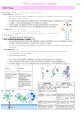
Hematoxylin staining Eosin staining Hematoxylin & Eosin staining
, MICROSCOPY
• Histology- Study of tissues and arrangement into organs
• Tissue = cells + extracellular matrix (ECM)
▪ Cells
– Major compartments = nucleus & cytoplasm(with organelles)
▪ ECM
– Structural network surrounding cells
– Influences cellular communication and function
– Contain fibres (collagen/elastic) and ground substance (proteoglycans, glycoproteins and
glycosaminoglycans)
• Structure is related to function!
HISTOLOGICAL TECHNIQUES
→ The routinely prepared hematoxylin and eosin–stained
section is the specimen most commonly studied.
• Haematoxylin
– Basic dye
– Stains acidic structures basophilic
– Colour: purple/dark blue
– Basic dyes react with anionic components of cells and tissue (components that carry a net negative
charge).
– A basic dye carries a net positive charge on its colored portion and is described by the general
formula [dye+ Cl− ].
– Anionic components include the phosphate groups of nucleic acids, the sulfate groups of
glycosaminoglycans, and the carboxyl groups of proteins.
– The ability of such anionic groups to react with a basic dye is called basophilia
– Hematoxylin is not, strictly speaking, a basic dye. It is used with a mordant (i.e., an intermediate link
between the tissue component and the dye).
– Tissue components that stain with hematoxylin also exhibit basophilia.
• Eosin
– Acidic dye
– Stains acidic structures eosinophilic
– Colour: pink
– An acidic dye carries a net negative charge on its colored portion and is described by the general
formula [Na+ dye−].
– Acidic dyes react with cationic groups in cells and tissues,
particularly with the ionized amino groups of proteins.
– The reaction of cationic groups with an acidic dye is
called acidophilia.
– Reactions of cell and tissue components with acidic
dyes are neither as specific nor as precise as reactions
with basic dyes
,→ A limited number of substances within cells and the extracellular matrix display basophilia.
• heterochromatin and nucleoli of the nucleus (chiefly because of ionized phosphate groups in nucleic
acids of both)
• cytoplasmic components such as the ergastoplasm (also because of ionized phosphate groups in
ribosomal RNA)
• extracellular materials such as the complex carbohydrates of the matrix of cartilage (because of
ionized sulfate groups).
→ Staining with acidic dyes is less specific, but more substances within cells and the extracellular matrix
exhibit acidophilia.
• most cytoplasmic filaments, especially those of muscle cells
• most intracellular membranous components and much of the otherwise unspecialized cytoplasm
• most extracellular fibers (primarily because of ionized amino groups).
→ Fixation, usually by a chemical or mixture of chemicals, permanently preserves the tissue structure for
subsequent treatments.
→ Specimens should be immersed in fixative immediately after they are removed from the body.
→ Fixation is used to:
• terminate cell metabolism,
• prevent enzymatic degradation of cells and tissues by autolysis (self-digestion),
• kill pathogenic microorganisms such as bacteria, fungi, and viruses
• harden the tissue as a result of either cross-linking or denaturing protein molecules.
Hematoxylin staining Eosin staining Hematoxylin & Eosin staining
, MICROSCOPY