(!#^(**#&#^&!#^&$*$1
*(!#^(**#&#^&!#^&$*$1
11/9/2026*(!#^(**#&#^&!#^&$*$1
of 125Practicode
of 125Practicode
Cases
of Cases
125Practicode
Part 211/9/2026,
Part 211/9/2026,
Cases
1:31:57
Part
1:31:57
211/9/2026,
AM11/9/2026Practicode
AM11/9/2026Practicode
1:31:57 AM11/9/2026Practicode
CasesCases
Part 2.pdfPage
Part 2.pdfPage
Cases
1 of 125@!*&#^&$*^@$^*&^$^$^*&^#%$*&^
Part
1 of 2.pdfPage
125@!*&#^&$*^@$^*&^$^$^*&^#%$*&^
1 of 125@!*&#^&$*^@$^*&^$^$^
Practicode Cases Part 2
Study online at https://quizlet.com/_ilp6df
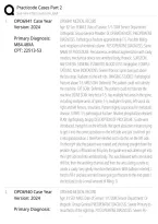
1. OPD6941 Case Year OPD6941 MEDICAL RECORD
Version: 2024 Age: 82 Sex: FEMALE Date of Service: 1/1/20XX Service Department:
Orthopedic Group General Provider: Dr. OPERATIVE NOTE: PREOPERATIVE
Primary Diagnosis: DIAGNOSIS: Pathological fracture approximately T-5. Possible Malig-
M84.48XA nant neoplasm of vertebral column. POSTOPERATIVE DIAGNOSIS: Same
CPT: 22513-53 NAME OF PROCEDURE: Percutaneous vertebral augmentation with cavity
creation, mechanical device one vertebral body, thoracic. SURGEON:
ANESTHESIA: GENERAL ESTIMATED BLOOD LOSS: Negligible. COMPLI-
CATIONS: None INDICATIONS: Severe thoracic spine pain just above
the bra strap. Radiates to the left ribs. IMAGING STUDIES: Pathological
fracture above T-5. MRI SCAN: Deferred. The patient could not tolerate
the machine. CAT SCAN: Deferred. The patient could not tolerate the
machine. BONE SCAN: Very hot at T-5. Has multiple hot areas in the spine,
including multiple areas of spine, T-5, multiple rib spots, left sacral ala,
right and left femurs, shoulders. Pattern highly suspicious for metastatic
disease. X-RAYS: T-5 pathological fracture. Alkaline phosphatase elevated
PLAN: Kyphoplasty, biopsy DESCRIPTION OF PROCEDURE: Guide wire
introduced, trying this on the left side. We spent about ten minutes trying
to get it into the correct position on the left side and just could not get
it into good position. I therefore elected not to do this on the left side.
On the right side the patient was rotated and shooting straight down the
pedicle. Again, infiltration on this plan, the guide wire was able to get into
the right side and into vertebral body. This was followed with cannulated
drill bit, then the working channel and then the articulating curette to
create a cavity. Very gently insertion the balloon. With balloon inserted, I
tried to fill it and was worried it was going to fracture to the end-plate. I
elected just to do a small amount of filling. O
2. OPD6940 Case Year OPD6940 MEDICAL RECORD
Version: 2024 Age: 81 SEX: MALE Date of Service: 1/1/20XX Service Department: Or-
thopedic Group General PREOPERATIVE DIAGNOSIS: Severe Primary os-
Primary Diagnosis: teoarthritis of the right hip. POSTOPERATIVE DIAGNOSIS: Severe Pri-
(!#^(**#&#^&!#^&$*$1
*(!#^(**#&#^&!#^&$*$1
*(!#^(**#&#^&!#^&$*$1
of 125Practicode
of 125Practicode
of 125Practicode
CasesCases
PartCases
211/9/2026,
Part 211/9/2026,
Part 211/9/2026,
1:31:57
1:31:57
AM11/9/2026Practicode
1:31:57
AM11/9/2026Practicode
AM11/9/2026Practicode
CasesCases
PartCases
2.pdfPage
Part 2.pdfPage
Part 2.pdfPage
1 of 125@!*&#^&$*^@$^*&^$^$^*&^#%$*&^
1 of 125@!*&#^&$*^@$^*&^$^$^*&^#%$*&^
1 of 125@!*&#^&$*^@$^*&^$^$^*&^#%$*&
,(!#^(**#&#^&!#^&$*$2
*(!#^(**#&#^&!#^&$*$2
11/9/2026*(!#^(**#&#^&!#^&$*$2
of 125Practicode
of 125Practicode
Cases
of Cases
125Practicode
Part 211/9/2026,
Part 211/9/2026,
Cases
1:31:57
Part
1:31:57
211/9/2026,
AM11/9/2026Practicode
AM11/9/2026Practicode
1:31:57 AM11/9/2026Practicode
CasesCases
Part 2.pdfPage
Part 2.pdfPage
Cases
2 of 125@!*&#^&$*^@$^*&^$^$^*&^#%$*&^
Part
2 of 2.pdfPage
125@!*&#^&$*^@$^*&^$^$^*&^#%$*&^
2 of 125@!*&#^&$*^@$^*&^$^$^
Practicode Cases Part 2
Study online at https://quizlet.com/_ilp6df
M16.11 mary osteoarthritis of the right hip. NAME OF PROCEDURE: Right AML
CPT: 27130-RT press-fit metal on metal total hip arthroplasty. SURGEON: ASSISTANT:
DESCRIPTION OF PROCEDURE: The patient was given 2 grams of Ancef
IV, a spinal anesthetic, then a general anesthetic for the procedure. Foley
catheter was placed in the bladder. The patient was placed in the left
lateral position where his right hip and lower extremity were prepped
and draped in the usual sterile fashion. Space suits were used. A straight
lateral approach was made and carefully carried down to the fascia
lata, which was split, bleeders were cauterized, a Charnley retractor was
placed. The anterior one-half of the gluteus medius and minimus free
from the greater trochanter, capsule was opened anterolaterally in a
T-shaped fashion. The hip was dislocated and the neck was sectioned.
Appropriate level for the AML component, the acetabular labrum was
excised, irrigated with PB solution and deepened and reamed to a 52
outside diameter. The wound was thoroughly irrigated with PB solution.
The spike pore-coated 52 outside diameter AML cup was impacted in 15
degrees of anteversion. A hole eliminator was applied. The wound was
irrigated with PB solution. A 36 inside diameter metal on metal compo-
nent was impacted. Attention was turned to the proximal femur, which
was prepared with reamers and broaches to accept the standard 10.5.
The wound was again irrigated with PB solution and the standard 10.5
fully coated prosthesis was impacted in neutral position. Trial reduction
proved a +12 gave excellent stability and leg length, therefore, the +12,
36 headed component was
3. OPD6938 Case Year OPD6938 MEDICAL RECORD
Version: 2022 Age: 57 SEX: FEMALE Date of Service: 1/1/20XX Service Department:
Orthopedic Group General Provider: OPERATIVE DIAGNOSIS: Right knee
Primary Diagnosis: symptomatic medial plica, with medial femoral condyle chondromala-
M94.261 cia. NAME OF PROCEDURE: Right knee examination under anesthesia,
Secondary Diagnosis: arthroscopy, medial plica excision, and medial femoral condyle chon-
droplasty. SURGEON: ASSISTANT: ANESTHESIA: General DESCRIPTION OF
(!#^(**#&#^&!#^&$*$2
*(!#^(**#&#^&!#^&$*$2
*(!#^(**#&#^&!#^&$*$2
of 125Practicode
of 125Practicode
of 125Practicode
CasesCases
PartCases
211/9/2026,
Part 211/9/2026,
Part 211/9/2026,
1:31:57
1:31:57
AM11/9/2026Practicode
1:31:57
AM11/9/2026Practicode
AM11/9/2026Practicode
CasesCases
PartCases
2.pdfPage
Part 2.pdfPage
Part 2.pdfPage
2 of 125@!*&#^&$*^@$^*&^$^$^*&^#%$*&^
2 of 125@!*&#^&$*^@$^*&^$^$^*&^#%$*&^
2 of 125@!*&#^&$*^@$^*&^$^$^*&^#%$*&
,(!#^(**#&#^&!#^&$*$3
*(!#^(**#&#^&!#^&$*$3
11/9/2026*(!#^(**#&#^&!#^&$*$3
of 125Practicode
of 125Practicode
Cases
of Cases
125Practicode
Part 211/9/2026,
Part 211/9/2026,
Cases
1:31:57
Part
1:31:57
211/9/2026,
AM11/9/2026Practicode
AM11/9/2026Practicode
1:31:57 AM11/9/2026Practicode
CasesCases
Part 2.pdfPage
Part 2.pdfPage
Cases
3 of 125@!*&#^&$*^@$^*&^$^$^*&^#%$*&^
Part
3 of 2.pdfPage
125@!*&#^&$*^@$^*&^$^$^*&^#%$*&^
3 of 125@!*&#^&$*^@$^*&^$^$^
Practicode Cases Part 2
Study online at https://quizlet.com/_ilp6df
M67.51 PROCEDURE: With the patient in the supine position under endotracheal
CPT: 29875-RT intubation with general anesthesia, the right knee was examined. She
had full range of motion, with all ligaments stable. No effusion. Portals
were preinjected. The knee was prepped and draped free in the usual
manner. Portals were established inferolaterally and inferomedially. In
the medial compartment, we identified normal cartilage on both articular
surfaces. Careful visualization and probing of the medial meniscus, both
superior and inferior surfaces, failed to reveal any sort of tearing. In the
notch, the anterior and posterior cruciate ligaments were intact to visual-
ization and probing. We did notice, however, that there was a large medi-
al plica present. In the lateral compartment, we found intact cartilage on
both articular surfaces. The lateral meniscus, popliteal tendon, and hiatus
were all intact to visualization and probing. In the patellofemoral joint,
we had normal alignment. Both the right patellar surface and femoral
trochlea were intact. However, in the medial gutter, we noted a very large
plica. There was tissue pinching between the patella and femur, and there
was chondromalacia over the edge of the femur. Therefore, we felt that
the meniscal tears that we suspected were not present and that this plica
could explain her symptoms. We therefore went ahead with excision of
the plica and a chondroplasty. We irrigated with st
4. OPD6998 Case Year OPD6998 MEDICAL RECORD
Version: 2022 OPERATIVE NOTE AGE: 35 SEX: Male DOS: 1/1/20XX PHYSICIAN: MD PRE-
OPERATIVE DIAGNOSIS: Biliary colic. POSTOPERATIVE DIAGNOSIS: Biliary
Primary Diagnosis: colic. OPERATIVE PROCEDURE: Laparoscopic cholecystectomy. SURGEON:
K80.50 MD ANESTHESIA: General and local. COMPLICATIONS: None. FINDINGS:
CPT: 47562 He had, of course, the normal-appearing gallbladder. There was no evi-
dence of inguinal herniation or other diseases. INDICATIONS: The patient
is a male who recently had a HIDA scan demonstrating 0% ejection
fraction. He has been having severe upper abdominal pain. He now
presents for cholecystectomy. DESCRIPTION OF PROCEDURE: After in-
formed consent was obtained, the patient was brought back to the oper-
(!#^(**#&#^&!#^&$*$3
*(!#^(**#&#^&!#^&$*$3
*(!#^(**#&#^&!#^&$*$3
of 125Practicode
of 125Practicode
of 125Practicode
CasesCases
PartCases
211/9/2026,
Part 211/9/2026,
Part 211/9/2026,
1:31:57
1:31:57
AM11/9/2026Practicode
1:31:57
AM11/9/2026Practicode
AM11/9/2026Practicode
CasesCases
PartCases
2.pdfPage
Part 2.pdfPage
Part 2.pdfPage
3 of 125@!*&#^&$*^@$^*&^$^$^*&^#%$*&^
3 of 125@!*&#^&$*^@$^*&^$^$^*&^#%$*&^
3 of 125@!*&#^&$*^@$^*&^$^$^*&^#%$*&
,(!#^(**#&#^&!#^&$*$4
*(!#^(**#&#^&!#^&$*$4
11/9/2026*(!#^(**#&#^&!#^&$*$4
of 125Practicode
of 125Practicode
Cases
of Cases
125Practicode
Part 211/9/2026,
Part 211/9/2026,
Cases
1:31:57
Part
1:31:57
211/9/2026,
AM11/9/2026Practicode
AM11/9/2026Practicode
1:31:57 AM11/9/2026Practicode
CasesCases
Part 2.pdfPage
Part 2.pdfPage
Cases
4 of 125@!*&#^&$*^@$^*&^$^$^*&^#%$*&^
Part
4 of 2.pdfPage
125@!*&#^&$*^@$^*&^$^$^*&^#%$*&^
4 of 125@!*&#^&$*^@$^*&^$^$^
Practicode Cases Part 2
Study online at https://quizlet.com/_ilp6df
ating room, placed on operating table in supine fashion. After adequate
monitors were placed, the patient was endotracheally intubated and
anesthetized. Compression boots were placed. The patient's abdomen
was prepped with Hibiclens soap and sterilely draped. A time-out was
performed, confirming the patient and the procedure. Local anesthetic
was infiltrated into the infraumbilical skin crease and the skin incision
was made. A Veress needle was passed into the peritoneal cavity without
difficulty and this was then inflated to a pressure of 15 mmHg using
carbon dioxide. A 5-mm Optiport was then positioned, demonstrating
appropriate placement. The laparoscope was then positioned and quick
visualization demonstrated no obvious abnormalities. There was no evi-
dence of inguinal hernias. Under direct vision, a 5-mm port was placed
in the right side of the abdomen and an 11-mm port in the subxiphoid
position. The patient was placed in reverse Trendelenburg and tilted to
the left side. The gallbladder was grasped and pushed up towards the
right hemidiaphragm. I was able to slowly free up the infundibulum and
the cystic duct and cystic artery. I con
5. OPD6982 Case Year OPD6982 MEDICAL RECORD
Version: 2024 AGE: 53 SEX: MALE DATE OF OPERATION:1/1/20XX PREOPERATIVE
DIAGNOSIS: ANAL CONDYLOMATAS PROCEDURES: FULGURATION OF
Primary Diagnosis: ANAL CONDYLOMATAS - ANOSCOPY POSTOPERATIVE DIAGNOSIS: ANAL
A63.0 CONDYLOMATAS SURGEON: MD Conscious Sedation: Intraservice Time
Secondary Diagnosis: 20 min. ESTIMATED BLOOD LOSS: MINIMAL. INDICATION: The patient
Z21 is a 53-year-old male positive for HIV. The patient came into OR for
CPT: 46612 anal condylomas. PROCEDURE: The patient was brought into the OR at 9
o'clock. The patient was put in prone position and procedure was done
under local anesthesia and conscious sedation. The patient was put in
prone position. The anal area was painted with Betadine and standard
drapes were placed around the area. After giving 16 ccs of lidocaine
IV, the anoscope was inserted in the anus and the electrocauterization
was used to remove the condylomas. The whole process took about
(!#^(**#&#^&!#^&$*$4
*(!#^(**#&#^&!#^&$*$4
*(!#^(**#&#^&!#^&$*$4
of 125Practicode
of 125Practicode
of 125Practicode
CasesCases
PartCases
211/9/2026,
Part 211/9/2026,
Part 211/9/2026,
1:31:57
1:31:57
AM11/9/2026Practicode
1:31:57
AM11/9/2026Practicode
AM11/9/2026Practicode
CasesCases
PartCases
2.pdfPage
Part 2.pdfPage
Part 2.pdfPage
4 of 125@!*&#^&$*^@$^*&^$^$^*&^#%$*&^
4 of 125@!*&#^&$*^@$^*&^$^$^*&^#%$*&^
4 of 125@!*&#^&$*^@$^*&^$^$^*&^#%$*&
*(!#^(**#&#^&!#^&$*$1
11/9/2026*(!#^(**#&#^&!#^&$*$1
of 125Practicode
of 125Practicode
Cases
of Cases
125Practicode
Part 211/9/2026,
Part 211/9/2026,
Cases
1:31:57
Part
1:31:57
211/9/2026,
AM11/9/2026Practicode
AM11/9/2026Practicode
1:31:57 AM11/9/2026Practicode
CasesCases
Part 2.pdfPage
Part 2.pdfPage
Cases
1 of 125@!*&#^&$*^@$^*&^$^$^*&^#%$*&^
Part
1 of 2.pdfPage
125@!*&#^&$*^@$^*&^$^$^*&^#%$*&^
1 of 125@!*&#^&$*^@$^*&^$^$^
Practicode Cases Part 2
Study online at https://quizlet.com/_ilp6df
1. OPD6941 Case Year OPD6941 MEDICAL RECORD
Version: 2024 Age: 82 Sex: FEMALE Date of Service: 1/1/20XX Service Department:
Orthopedic Group General Provider: Dr. OPERATIVE NOTE: PREOPERATIVE
Primary Diagnosis: DIAGNOSIS: Pathological fracture approximately T-5. Possible Malig-
M84.48XA nant neoplasm of vertebral column. POSTOPERATIVE DIAGNOSIS: Same
CPT: 22513-53 NAME OF PROCEDURE: Percutaneous vertebral augmentation with cavity
creation, mechanical device one vertebral body, thoracic. SURGEON:
ANESTHESIA: GENERAL ESTIMATED BLOOD LOSS: Negligible. COMPLI-
CATIONS: None INDICATIONS: Severe thoracic spine pain just above
the bra strap. Radiates to the left ribs. IMAGING STUDIES: Pathological
fracture above T-5. MRI SCAN: Deferred. The patient could not tolerate
the machine. CAT SCAN: Deferred. The patient could not tolerate the
machine. BONE SCAN: Very hot at T-5. Has multiple hot areas in the spine,
including multiple areas of spine, T-5, multiple rib spots, left sacral ala,
right and left femurs, shoulders. Pattern highly suspicious for metastatic
disease. X-RAYS: T-5 pathological fracture. Alkaline phosphatase elevated
PLAN: Kyphoplasty, biopsy DESCRIPTION OF PROCEDURE: Guide wire
introduced, trying this on the left side. We spent about ten minutes trying
to get it into the correct position on the left side and just could not get
it into good position. I therefore elected not to do this on the left side.
On the right side the patient was rotated and shooting straight down the
pedicle. Again, infiltration on this plan, the guide wire was able to get into
the right side and into vertebral body. This was followed with cannulated
drill bit, then the working channel and then the articulating curette to
create a cavity. Very gently insertion the balloon. With balloon inserted, I
tried to fill it and was worried it was going to fracture to the end-plate. I
elected just to do a small amount of filling. O
2. OPD6940 Case Year OPD6940 MEDICAL RECORD
Version: 2024 Age: 81 SEX: MALE Date of Service: 1/1/20XX Service Department: Or-
thopedic Group General PREOPERATIVE DIAGNOSIS: Severe Primary os-
Primary Diagnosis: teoarthritis of the right hip. POSTOPERATIVE DIAGNOSIS: Severe Pri-
(!#^(**#&#^&!#^&$*$1
*(!#^(**#&#^&!#^&$*$1
*(!#^(**#&#^&!#^&$*$1
of 125Practicode
of 125Practicode
of 125Practicode
CasesCases
PartCases
211/9/2026,
Part 211/9/2026,
Part 211/9/2026,
1:31:57
1:31:57
AM11/9/2026Practicode
1:31:57
AM11/9/2026Practicode
AM11/9/2026Practicode
CasesCases
PartCases
2.pdfPage
Part 2.pdfPage
Part 2.pdfPage
1 of 125@!*&#^&$*^@$^*&^$^$^*&^#%$*&^
1 of 125@!*&#^&$*^@$^*&^$^$^*&^#%$*&^
1 of 125@!*&#^&$*^@$^*&^$^$^*&^#%$*&
,(!#^(**#&#^&!#^&$*$2
*(!#^(**#&#^&!#^&$*$2
11/9/2026*(!#^(**#&#^&!#^&$*$2
of 125Practicode
of 125Practicode
Cases
of Cases
125Practicode
Part 211/9/2026,
Part 211/9/2026,
Cases
1:31:57
Part
1:31:57
211/9/2026,
AM11/9/2026Practicode
AM11/9/2026Practicode
1:31:57 AM11/9/2026Practicode
CasesCases
Part 2.pdfPage
Part 2.pdfPage
Cases
2 of 125@!*&#^&$*^@$^*&^$^$^*&^#%$*&^
Part
2 of 2.pdfPage
125@!*&#^&$*^@$^*&^$^$^*&^#%$*&^
2 of 125@!*&#^&$*^@$^*&^$^$^
Practicode Cases Part 2
Study online at https://quizlet.com/_ilp6df
M16.11 mary osteoarthritis of the right hip. NAME OF PROCEDURE: Right AML
CPT: 27130-RT press-fit metal on metal total hip arthroplasty. SURGEON: ASSISTANT:
DESCRIPTION OF PROCEDURE: The patient was given 2 grams of Ancef
IV, a spinal anesthetic, then a general anesthetic for the procedure. Foley
catheter was placed in the bladder. The patient was placed in the left
lateral position where his right hip and lower extremity were prepped
and draped in the usual sterile fashion. Space suits were used. A straight
lateral approach was made and carefully carried down to the fascia
lata, which was split, bleeders were cauterized, a Charnley retractor was
placed. The anterior one-half of the gluteus medius and minimus free
from the greater trochanter, capsule was opened anterolaterally in a
T-shaped fashion. The hip was dislocated and the neck was sectioned.
Appropriate level for the AML component, the acetabular labrum was
excised, irrigated with PB solution and deepened and reamed to a 52
outside diameter. The wound was thoroughly irrigated with PB solution.
The spike pore-coated 52 outside diameter AML cup was impacted in 15
degrees of anteversion. A hole eliminator was applied. The wound was
irrigated with PB solution. A 36 inside diameter metal on metal compo-
nent was impacted. Attention was turned to the proximal femur, which
was prepared with reamers and broaches to accept the standard 10.5.
The wound was again irrigated with PB solution and the standard 10.5
fully coated prosthesis was impacted in neutral position. Trial reduction
proved a +12 gave excellent stability and leg length, therefore, the +12,
36 headed component was
3. OPD6938 Case Year OPD6938 MEDICAL RECORD
Version: 2022 Age: 57 SEX: FEMALE Date of Service: 1/1/20XX Service Department:
Orthopedic Group General Provider: OPERATIVE DIAGNOSIS: Right knee
Primary Diagnosis: symptomatic medial plica, with medial femoral condyle chondromala-
M94.261 cia. NAME OF PROCEDURE: Right knee examination under anesthesia,
Secondary Diagnosis: arthroscopy, medial plica excision, and medial femoral condyle chon-
droplasty. SURGEON: ASSISTANT: ANESTHESIA: General DESCRIPTION OF
(!#^(**#&#^&!#^&$*$2
*(!#^(**#&#^&!#^&$*$2
*(!#^(**#&#^&!#^&$*$2
of 125Practicode
of 125Practicode
of 125Practicode
CasesCases
PartCases
211/9/2026,
Part 211/9/2026,
Part 211/9/2026,
1:31:57
1:31:57
AM11/9/2026Practicode
1:31:57
AM11/9/2026Practicode
AM11/9/2026Practicode
CasesCases
PartCases
2.pdfPage
Part 2.pdfPage
Part 2.pdfPage
2 of 125@!*&#^&$*^@$^*&^$^$^*&^#%$*&^
2 of 125@!*&#^&$*^@$^*&^$^$^*&^#%$*&^
2 of 125@!*&#^&$*^@$^*&^$^$^*&^#%$*&
,(!#^(**#&#^&!#^&$*$3
*(!#^(**#&#^&!#^&$*$3
11/9/2026*(!#^(**#&#^&!#^&$*$3
of 125Practicode
of 125Practicode
Cases
of Cases
125Practicode
Part 211/9/2026,
Part 211/9/2026,
Cases
1:31:57
Part
1:31:57
211/9/2026,
AM11/9/2026Practicode
AM11/9/2026Practicode
1:31:57 AM11/9/2026Practicode
CasesCases
Part 2.pdfPage
Part 2.pdfPage
Cases
3 of 125@!*&#^&$*^@$^*&^$^$^*&^#%$*&^
Part
3 of 2.pdfPage
125@!*&#^&$*^@$^*&^$^$^*&^#%$*&^
3 of 125@!*&#^&$*^@$^*&^$^$^
Practicode Cases Part 2
Study online at https://quizlet.com/_ilp6df
M67.51 PROCEDURE: With the patient in the supine position under endotracheal
CPT: 29875-RT intubation with general anesthesia, the right knee was examined. She
had full range of motion, with all ligaments stable. No effusion. Portals
were preinjected. The knee was prepped and draped free in the usual
manner. Portals were established inferolaterally and inferomedially. In
the medial compartment, we identified normal cartilage on both articular
surfaces. Careful visualization and probing of the medial meniscus, both
superior and inferior surfaces, failed to reveal any sort of tearing. In the
notch, the anterior and posterior cruciate ligaments were intact to visual-
ization and probing. We did notice, however, that there was a large medi-
al plica present. In the lateral compartment, we found intact cartilage on
both articular surfaces. The lateral meniscus, popliteal tendon, and hiatus
were all intact to visualization and probing. In the patellofemoral joint,
we had normal alignment. Both the right patellar surface and femoral
trochlea were intact. However, in the medial gutter, we noted a very large
plica. There was tissue pinching between the patella and femur, and there
was chondromalacia over the edge of the femur. Therefore, we felt that
the meniscal tears that we suspected were not present and that this plica
could explain her symptoms. We therefore went ahead with excision of
the plica and a chondroplasty. We irrigated with st
4. OPD6998 Case Year OPD6998 MEDICAL RECORD
Version: 2022 OPERATIVE NOTE AGE: 35 SEX: Male DOS: 1/1/20XX PHYSICIAN: MD PRE-
OPERATIVE DIAGNOSIS: Biliary colic. POSTOPERATIVE DIAGNOSIS: Biliary
Primary Diagnosis: colic. OPERATIVE PROCEDURE: Laparoscopic cholecystectomy. SURGEON:
K80.50 MD ANESTHESIA: General and local. COMPLICATIONS: None. FINDINGS:
CPT: 47562 He had, of course, the normal-appearing gallbladder. There was no evi-
dence of inguinal herniation or other diseases. INDICATIONS: The patient
is a male who recently had a HIDA scan demonstrating 0% ejection
fraction. He has been having severe upper abdominal pain. He now
presents for cholecystectomy. DESCRIPTION OF PROCEDURE: After in-
formed consent was obtained, the patient was brought back to the oper-
(!#^(**#&#^&!#^&$*$3
*(!#^(**#&#^&!#^&$*$3
*(!#^(**#&#^&!#^&$*$3
of 125Practicode
of 125Practicode
of 125Practicode
CasesCases
PartCases
211/9/2026,
Part 211/9/2026,
Part 211/9/2026,
1:31:57
1:31:57
AM11/9/2026Practicode
1:31:57
AM11/9/2026Practicode
AM11/9/2026Practicode
CasesCases
PartCases
2.pdfPage
Part 2.pdfPage
Part 2.pdfPage
3 of 125@!*&#^&$*^@$^*&^$^$^*&^#%$*&^
3 of 125@!*&#^&$*^@$^*&^$^$^*&^#%$*&^
3 of 125@!*&#^&$*^@$^*&^$^$^*&^#%$*&
,(!#^(**#&#^&!#^&$*$4
*(!#^(**#&#^&!#^&$*$4
11/9/2026*(!#^(**#&#^&!#^&$*$4
of 125Practicode
of 125Practicode
Cases
of Cases
125Practicode
Part 211/9/2026,
Part 211/9/2026,
Cases
1:31:57
Part
1:31:57
211/9/2026,
AM11/9/2026Practicode
AM11/9/2026Practicode
1:31:57 AM11/9/2026Practicode
CasesCases
Part 2.pdfPage
Part 2.pdfPage
Cases
4 of 125@!*&#^&$*^@$^*&^$^$^*&^#%$*&^
Part
4 of 2.pdfPage
125@!*&#^&$*^@$^*&^$^$^*&^#%$*&^
4 of 125@!*&#^&$*^@$^*&^$^$^
Practicode Cases Part 2
Study online at https://quizlet.com/_ilp6df
ating room, placed on operating table in supine fashion. After adequate
monitors were placed, the patient was endotracheally intubated and
anesthetized. Compression boots were placed. The patient's abdomen
was prepped with Hibiclens soap and sterilely draped. A time-out was
performed, confirming the patient and the procedure. Local anesthetic
was infiltrated into the infraumbilical skin crease and the skin incision
was made. A Veress needle was passed into the peritoneal cavity without
difficulty and this was then inflated to a pressure of 15 mmHg using
carbon dioxide. A 5-mm Optiport was then positioned, demonstrating
appropriate placement. The laparoscope was then positioned and quick
visualization demonstrated no obvious abnormalities. There was no evi-
dence of inguinal hernias. Under direct vision, a 5-mm port was placed
in the right side of the abdomen and an 11-mm port in the subxiphoid
position. The patient was placed in reverse Trendelenburg and tilted to
the left side. The gallbladder was grasped and pushed up towards the
right hemidiaphragm. I was able to slowly free up the infundibulum and
the cystic duct and cystic artery. I con
5. OPD6982 Case Year OPD6982 MEDICAL RECORD
Version: 2024 AGE: 53 SEX: MALE DATE OF OPERATION:1/1/20XX PREOPERATIVE
DIAGNOSIS: ANAL CONDYLOMATAS PROCEDURES: FULGURATION OF
Primary Diagnosis: ANAL CONDYLOMATAS - ANOSCOPY POSTOPERATIVE DIAGNOSIS: ANAL
A63.0 CONDYLOMATAS SURGEON: MD Conscious Sedation: Intraservice Time
Secondary Diagnosis: 20 min. ESTIMATED BLOOD LOSS: MINIMAL. INDICATION: The patient
Z21 is a 53-year-old male positive for HIV. The patient came into OR for
CPT: 46612 anal condylomas. PROCEDURE: The patient was brought into the OR at 9
o'clock. The patient was put in prone position and procedure was done
under local anesthesia and conscious sedation. The patient was put in
prone position. The anal area was painted with Betadine and standard
drapes were placed around the area. After giving 16 ccs of lidocaine
IV, the anoscope was inserted in the anus and the electrocauterization
was used to remove the condylomas. The whole process took about
(!#^(**#&#^&!#^&$*$4
*(!#^(**#&#^&!#^&$*$4
*(!#^(**#&#^&!#^&$*$4
of 125Practicode
of 125Practicode
of 125Practicode
CasesCases
PartCases
211/9/2026,
Part 211/9/2026,
Part 211/9/2026,
1:31:57
1:31:57
AM11/9/2026Practicode
1:31:57
AM11/9/2026Practicode
AM11/9/2026Practicode
CasesCases
PartCases
2.pdfPage
Part 2.pdfPage
Part 2.pdfPage
4 of 125@!*&#^&$*^@$^*&^$^$^*&^#%$*&^
4 of 125@!*&#^&$*^@$^*&^$^$^*&^#%$*&^
4 of 125@!*&#^&$*^@$^*&^$^$^*&^#%$*&