
2026,
oot_tip.pdf10/16/2026,
5:42:03
PageAM*&!#%#&&!%%#^#^&^!#&*^&^*#!(FG*T!^&*#*((*!#&()#_)#!*&$!*#)(!*W.A_Lab_5.1__onion_root_tip.pdf10/16/2026,
1@~!@#########################Page
5:42:03 AM*&!#%#&&!%%#^#^&^!#&*^&^*#!(FG*T!^&*#*((*!#&()#_)#!*&$!*#)(!*##(*0W.A_Lab_5.1__onion_root_tip.pdf.pdf*&!#%#&&!%%#^#
1 of 910/16/2026, 5:42:03 AM*&!#%#&&!%%#^#^&^!#&*^&^*#!(FG*T!^&*#*((*!#&()#_)#!*&$!*#)(!*W.A_Lab_5.1__
5:42:03 AM*&!#%#&&!%%#^#^&^!#&*^&^
1
Walker Alejandro 11/3/23 Biology Dual Credit
Lab 5.1 Mitosis (cell division) 10/24/23 Total: 100 points
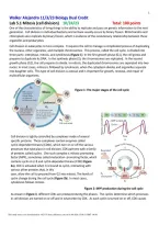
One of the characteristics of living things is the ability to replicate and pass on genetic information to the next
generation. Cell division in individual bacteria and archaea usually occurs by binary fission. Mitochondria and
chloroplasts also replicate by binary fission, which is evidence of the evolutionary relationship between these
organelles and prokaryotes.
Cell division in eukaryotes is more complex. It requires the cell to manage a complicated process of duplicating
the nucleus, other organelles, and multiple chromosomes. This process, called the cell cycle, is divided into
three parts: interphase, mitosis, and cytokinesis (Figure 1). In the first growth phase (G1), the cell grows and
prepares to duplicate its DNA. In the synthesis phase (S), the chromosomes are replicated. In the second
growth phase (G2), the cell prepares to divide. In mitosis, the duplicated chromosomes are separated into two
nuclei. In most cases, mitosis is followed by cytokinesis, when the cytoplasm divides and organelles separate
into daughter cells. This type of cell division is asexual and is important for growth, renewal, and repair of
multicellular organisms.
Figure 1: The major stages of the cell cycle
Cell division is tightly controlled by complexes made of several
specific proteins. These complexes contain enzymes called
cyclin-dependent kinases (CDKs), which turn on or off the various
processes that take place in cell division. CDK partners with a family
of proteins called cyclins. One such complex is mitosis-promoting
factor (MPF), sometimes called maturation- promoting factor, which
contains cyclin A or B and cyclin-dependent kinase (CDK) (Figure
2a). CDK is activated when it is bound to cyclin, interacting with
various other proteins that, in this
case, allow the cell to proceed from G2 into mitosis. The levels of
cyclin change during the cell cycle (Figure 2b). In most cases,
cytokinesis follows mitosis.
Figure 2: MPF production during the cell cycle
As shown in Figure 3, different CDKs are produced during the phases. The cyclins determine which processes
in cell division are turned on or off and in what order by CDK. As each cyclin is turned on or off, CDK causes
This study source was downloaded by 1827175 from cliffsnotes.com on 01-08-2026 15:00:33 GMT -06:00
df10/16/2026,
*&!#%#&&!%%#^#^&^!#&*^&^*#!(FG*T!^&*#*((*!#&()#_)#!*&$!*#)(!*W.A_Lab_5.1__onion_root_tip.pdf10/16/2026,
@~!@#########################Page
5:42:03 AM*&!#%#&&!%%#^#^&^!#&*^&^*#!(FG*T!^&*#*((*!#&()#_)#!*&$!*#)(!*##(*0W.A_Lab_5.1__onion_root_tip.pdf.pdf*&!#%#&&!%%#^#^&^!#&*^
1 of 910/16/2026, 5:42:03 AM*&!#%#&&!%%#^#^&^!#&*^&^*#!(FG*T!^&*#*((*!#&()#_)#!*&$!*#)(!*W.A_Lab_5.1__onion_r
https://www.cliffsnotes.com//study-notes/16937987 5:42:03 AM*&!#%#&&!%%#^#^&^!#&*^&^*#!(FG*T!^&*#*((*
,2026,
oot_tip.pdf10/16/2026,
5:42:03
PageAM*&!#%#&&!%%#^#^&^!#&*^&^*#!(FG*T!^&*#*((*!#&()#_)#!*&$!*#)(!*W.A_Lab_5.1__onion_root_tip.pdf10/16/2026,
2@~!@#########################Page
5:42:03 AM*&!#%#&&!%%#^#^&^!#&*^&^*#!(FG*T!^&*#*((*!#&()#_)#!*&$!*#)(!*##(*0W.A_Lab_5.1__onion_root_tip.pdf.pdf*&!#%#&&!%%#^#
2 of 910/16/2026, 5:42:03 AM*&!#%#&&!%%#^#^&^!#&*^&^*#!(FG*T!^&*#*((*!#&()#_)#!*&$!*#)(!*W.A_Lab_5.1__
5:42:03 AM*&!#%#&&!%%#^#^&^!#&*^&^
2
the cell to progress through the stages in the cell cycle.
Figure 3: Levels of CDKs
during the cell cycle
Cyclins and CDKs do not allow the cell to progress through its
cycle automatically. There are three checkpoints a cell must
pass through: the G1 checkpoint, G2 checkpoint, and the M-
spindle checkpoint (Figure 4). At each of the checkpoints, the
cell checks that it has completed all of the tasks needed and is
ready to proceed to the next step in its cycle. Cells pass the G1
checkpoint when they are stimulated by appropriate external
growth factors; for example, platelet-derived growth factor
(PDGF) stimulates cells near a wound to divide so that they
can repair the injury. The G2 checkpoint checks for damage
after DNA is replicated, and if there is damage, it prevents the
cell from going into mitosis. The M-spindle (metaphase)
checkpoint assures that the mitotic spindles or microtubules
are properly attached to the kinetochores (anchor sites on the
chromosomes). If the spindles are not anchored properly, the
cell does not continue on through mitosis. The cell cycle is
regulated very precisely.
Mutations in cell cycle genes that interfere with proper cell
cycle control are found very often in cancer cells.
Figure 1 illustrates the green onion (Allium) root tip. Because all
primary growth occurs in the apical meristem of plant roots and
shoots, we expect to see more cells growing and dividing in that
area.
This study source was downloaded by 1827175 from cliffsnotes.com on 01-08-2026 15:00:33 GMT -06:00
df10/16/2026,
*&!#%#&&!%%#^#^&^!#&*^&^*#!(FG*T!^&*#*((*!#&()#_)#!*&$!*#)(!*W.A_Lab_5.1__onion_root_tip.pdf10/16/2026,
@~!@#########################Page
5:42:03 AM*&!#%#&&!%%#^#^&^!#&*^&^*#!(FG*T!^&*#*((*!#&()#_)#!*&$!*#)(!*##(*0W.A_Lab_5.1__onion_root_tip.pdf.pdf*&!#%#&&!%%#^#^&^!#&*^
2 of 910/16/2026, 5:42:03 AM*&!#%#&&!%%#^#^&^!#&*^&^*#!(FG*T!^&*#*((*!#&()#_)#!*&$!*#)(!*W.A_Lab_5.1__onion_r
https://www.cliffsnotes.com//study-notes/16937987 5:42:03 AM*&!#%#&&!%%#^#^&^!#&*^&^*#!(FG*T!^&*#*((*
oot_tip.pdf10/16/2026,
5:42:03
PageAM*&!#%#&&!%%#^#^&^!#&*^&^*#!(FG*T!^&*#*((*!#&()#_)#!*&$!*#)(!*W.A_Lab_5.1__onion_root_tip.pdf10/16/2026,
1@~!@#########################Page
5:42:03 AM*&!#%#&&!%%#^#^&^!#&*^&^*#!(FG*T!^&*#*((*!#&()#_)#!*&$!*#)(!*##(*0W.A_Lab_5.1__onion_root_tip.pdf.pdf*&!#%#&&!%%#^#
1 of 910/16/2026, 5:42:03 AM*&!#%#&&!%%#^#^&^!#&*^&^*#!(FG*T!^&*#*((*!#&()#_)#!*&$!*#)(!*W.A_Lab_5.1__
5:42:03 AM*&!#%#&&!%%#^#^&^!#&*^&^
1
Walker Alejandro 11/3/23 Biology Dual Credit
Lab 5.1 Mitosis (cell division) 10/24/23 Total: 100 points
One of the characteristics of living things is the ability to replicate and pass on genetic information to the next
generation. Cell division in individual bacteria and archaea usually occurs by binary fission. Mitochondria and
chloroplasts also replicate by binary fission, which is evidence of the evolutionary relationship between these
organelles and prokaryotes.
Cell division in eukaryotes is more complex. It requires the cell to manage a complicated process of duplicating
the nucleus, other organelles, and multiple chromosomes. This process, called the cell cycle, is divided into
three parts: interphase, mitosis, and cytokinesis (Figure 1). In the first growth phase (G1), the cell grows and
prepares to duplicate its DNA. In the synthesis phase (S), the chromosomes are replicated. In the second
growth phase (G2), the cell prepares to divide. In mitosis, the duplicated chromosomes are separated into two
nuclei. In most cases, mitosis is followed by cytokinesis, when the cytoplasm divides and organelles separate
into daughter cells. This type of cell division is asexual and is important for growth, renewal, and repair of
multicellular organisms.
Figure 1: The major stages of the cell cycle
Cell division is tightly controlled by complexes made of several
specific proteins. These complexes contain enzymes called
cyclin-dependent kinases (CDKs), which turn on or off the various
processes that take place in cell division. CDK partners with a family
of proteins called cyclins. One such complex is mitosis-promoting
factor (MPF), sometimes called maturation- promoting factor, which
contains cyclin A or B and cyclin-dependent kinase (CDK) (Figure
2a). CDK is activated when it is bound to cyclin, interacting with
various other proteins that, in this
case, allow the cell to proceed from G2 into mitosis. The levels of
cyclin change during the cell cycle (Figure 2b). In most cases,
cytokinesis follows mitosis.
Figure 2: MPF production during the cell cycle
As shown in Figure 3, different CDKs are produced during the phases. The cyclins determine which processes
in cell division are turned on or off and in what order by CDK. As each cyclin is turned on or off, CDK causes
This study source was downloaded by 1827175 from cliffsnotes.com on 01-08-2026 15:00:33 GMT -06:00
df10/16/2026,
*&!#%#&&!%%#^#^&^!#&*^&^*#!(FG*T!^&*#*((*!#&()#_)#!*&$!*#)(!*W.A_Lab_5.1__onion_root_tip.pdf10/16/2026,
@~!@#########################Page
5:42:03 AM*&!#%#&&!%%#^#^&^!#&*^&^*#!(FG*T!^&*#*((*!#&()#_)#!*&$!*#)(!*##(*0W.A_Lab_5.1__onion_root_tip.pdf.pdf*&!#%#&&!%%#^#^&^!#&*^
1 of 910/16/2026, 5:42:03 AM*&!#%#&&!%%#^#^&^!#&*^&^*#!(FG*T!^&*#*((*!#&()#_)#!*&$!*#)(!*W.A_Lab_5.1__onion_r
https://www.cliffsnotes.com//study-notes/16937987 5:42:03 AM*&!#%#&&!%%#^#^&^!#&*^&^*#!(FG*T!^&*#*((*
,2026,
oot_tip.pdf10/16/2026,
5:42:03
PageAM*&!#%#&&!%%#^#^&^!#&*^&^*#!(FG*T!^&*#*((*!#&()#_)#!*&$!*#)(!*W.A_Lab_5.1__onion_root_tip.pdf10/16/2026,
2@~!@#########################Page
5:42:03 AM*&!#%#&&!%%#^#^&^!#&*^&^*#!(FG*T!^&*#*((*!#&()#_)#!*&$!*#)(!*##(*0W.A_Lab_5.1__onion_root_tip.pdf.pdf*&!#%#&&!%%#^#
2 of 910/16/2026, 5:42:03 AM*&!#%#&&!%%#^#^&^!#&*^&^*#!(FG*T!^&*#*((*!#&()#_)#!*&$!*#)(!*W.A_Lab_5.1__
5:42:03 AM*&!#%#&&!%%#^#^&^!#&*^&^
2
the cell to progress through the stages in the cell cycle.
Figure 3: Levels of CDKs
during the cell cycle
Cyclins and CDKs do not allow the cell to progress through its
cycle automatically. There are three checkpoints a cell must
pass through: the G1 checkpoint, G2 checkpoint, and the M-
spindle checkpoint (Figure 4). At each of the checkpoints, the
cell checks that it has completed all of the tasks needed and is
ready to proceed to the next step in its cycle. Cells pass the G1
checkpoint when they are stimulated by appropriate external
growth factors; for example, platelet-derived growth factor
(PDGF) stimulates cells near a wound to divide so that they
can repair the injury. The G2 checkpoint checks for damage
after DNA is replicated, and if there is damage, it prevents the
cell from going into mitosis. The M-spindle (metaphase)
checkpoint assures that the mitotic spindles or microtubules
are properly attached to the kinetochores (anchor sites on the
chromosomes). If the spindles are not anchored properly, the
cell does not continue on through mitosis. The cell cycle is
regulated very precisely.
Mutations in cell cycle genes that interfere with proper cell
cycle control are found very often in cancer cells.
Figure 1 illustrates the green onion (Allium) root tip. Because all
primary growth occurs in the apical meristem of plant roots and
shoots, we expect to see more cells growing and dividing in that
area.
This study source was downloaded by 1827175 from cliffsnotes.com on 01-08-2026 15:00:33 GMT -06:00
df10/16/2026,
*&!#%#&&!%%#^#^&^!#&*^&^*#!(FG*T!^&*#*((*!#&()#_)#!*&$!*#)(!*W.A_Lab_5.1__onion_root_tip.pdf10/16/2026,
@~!@#########################Page
5:42:03 AM*&!#%#&&!%%#^#^&^!#&*^&^*#!(FG*T!^&*#*((*!#&()#_)#!*&$!*#)(!*##(*0W.A_Lab_5.1__onion_root_tip.pdf.pdf*&!#%#&&!%%#^#^&^!#&*^
2 of 910/16/2026, 5:42:03 AM*&!#%#&&!%%#^#^&^!#&*^&^*#!(FG*T!^&*#*((*!#&()#_)#!*&$!*#)(!*W.A_Lab_5.1__onion_r
https://www.cliffsnotes.com//study-notes/16937987 5:42:03 AM*&!#%#&&!%%#^#^&^!#&*^&^*#!(FG*T!^&*#*((*


