
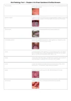
Oral Pathology Test 1 - Chapters 3 & 4 Exam Questions & Verified Answers
filiform papillae smallest and most numerous structure on the tongue. They are mechanical and DO
NOT contain tastebuds.
fungiform papillae brighter red, raised, mushroom shaped containing tastebuds. Fewer in number than
filiform, most often located on the lateral boarder and anterior tip of the tongue
foliate papillae arranged in the vertical folds of the tongue containing taste buds.
circumvallate papillae largest of the papillae and appear as 2 - 4mm papules in a v shaped row in the
posterior aspect of the dorsum of the tongue
macule a circumscribed area of the epidermis or mucosa distinguished by color different
from surrounding tissue. It is flat, not raised above the surface and may appear
alone or in groups and may be blue, brown, black, or red
papule circumscribed, often inflammatory, elevated lesions, protruding above surface of
surrounding tissue. A solid lesion that is less than 1cm in diameter.
nodule a solid mass of tissue that has the dimension of depth (extends deep into the
dermis) up to 1 cm in diameter. Usually detected by palpation
sessile describing the base of a lesion that is flat, attached directly by the base.
pedunculated attached by a stem like or stalk base similar to that of a mushroom
, Oral Pathology Test 1 - Chapters 3 & 4 Exam Questions & Verified Answers
papillary a small, nipple-shaped projection or elevation that may be found in clusters
vesicle a circumscribed, fluid-filled elevation that is less than 1 cm in diameter. The fluid
consists of serous fluid but may contain blood and infectious agents. The epithelial
lining is thin and will eventually rupture causing an ulcer
bulla the diameter of the circumscribed, elevated lesion exceeds 5mm. This is larger than
a vesicle and usually contains serous fluid and looks like a blister.
lobule a segment of lobe that is a part of the whole. They may be found fused together
pustule a circumscribed elevation filled with purulent exudate resulting from an infection.
Less than 1 cm in diameter and may be preceded by a vesicle or papule
ulcer an uncovered wound of cutaneous or mucosal tissue that exhibits gradual tissue
disintegration and necrosis. This extends deeper than erosion, form beyond the
basal layer of the epithelium into the dermis. Usually painful - topical or systemic
drug therapy may be required for management.
erosion a lesion where the epithelium above the basal layer has eroded. They are moist and
slightly depressed resulting from a broken vesicle or trauma. No scaring is involved
, Oral Pathology Test 1 - Chapters 3 & 4 Exam Questions & Verified Answers
plaque a flat, solid ,raised area that is larger than 1cm in diameter. Usually superficial but
can extend into the dermis. Edges may be sloped and sometimes surface keratin
can exist. ei) leukoplakia
fissure normal or abnormal linear cleft in the epidermis. May affect the tongue, lips and
perioral tissues. Disease may occur when a pathogenic organism infects the area
causing pain, ulceration, and inflammation
cyst located in the dermis, subcutaneous tissue or bone. This results from entrapment of
fluids, gas, or semi solid substances that grow to produce an enclosed capsule or
cavity. Contains clear fluid that appears pink to blue whereas keratin-filled appear
yellow or creamy
tumor a solid mass of tissue larger than 1 cm in diameter and also had the dimension of
depth. Term also used to represent a neoplasm. Classified into benign or malignant.
Benign grow more slowly and less aggressive and appear raised and have defined
borders. Malignant usually aggressive and spread rapidly and have ill defined
borders - have the ability to metastasize
exophytic lesion growing outward from the surface of a tissue is known as?
endophytic lesion growing into the surrounding tissues which present as palpable masses with
or without noticeable swelling.
erythema abnormal redness or the mucosa or gingiva
pallor paleness of skin or mucosal tissue
blanching red to white upon pressure
filiform papillae smallest and most numerous structure on the tongue. They are mechanical and DO
NOT contain tastebuds.
fungiform papillae brighter red, raised, mushroom shaped containing tastebuds. Fewer in number than
filiform, most often located on the lateral boarder and anterior tip of the tongue
foliate papillae arranged in the vertical folds of the tongue containing taste buds.
circumvallate papillae largest of the papillae and appear as 2 - 4mm papules in a v shaped row in the
posterior aspect of the dorsum of the tongue
macule a circumscribed area of the epidermis or mucosa distinguished by color different
from surrounding tissue. It is flat, not raised above the surface and may appear
alone or in groups and may be blue, brown, black, or red
papule circumscribed, often inflammatory, elevated lesions, protruding above surface of
surrounding tissue. A solid lesion that is less than 1cm in diameter.
nodule a solid mass of tissue that has the dimension of depth (extends deep into the
dermis) up to 1 cm in diameter. Usually detected by palpation
sessile describing the base of a lesion that is flat, attached directly by the base.
pedunculated attached by a stem like or stalk base similar to that of a mushroom
, Oral Pathology Test 1 - Chapters 3 & 4 Exam Questions & Verified Answers
papillary a small, nipple-shaped projection or elevation that may be found in clusters
vesicle a circumscribed, fluid-filled elevation that is less than 1 cm in diameter. The fluid
consists of serous fluid but may contain blood and infectious agents. The epithelial
lining is thin and will eventually rupture causing an ulcer
bulla the diameter of the circumscribed, elevated lesion exceeds 5mm. This is larger than
a vesicle and usually contains serous fluid and looks like a blister.
lobule a segment of lobe that is a part of the whole. They may be found fused together
pustule a circumscribed elevation filled with purulent exudate resulting from an infection.
Less than 1 cm in diameter and may be preceded by a vesicle or papule
ulcer an uncovered wound of cutaneous or mucosal tissue that exhibits gradual tissue
disintegration and necrosis. This extends deeper than erosion, form beyond the
basal layer of the epithelium into the dermis. Usually painful - topical or systemic
drug therapy may be required for management.
erosion a lesion where the epithelium above the basal layer has eroded. They are moist and
slightly depressed resulting from a broken vesicle or trauma. No scaring is involved
, Oral Pathology Test 1 - Chapters 3 & 4 Exam Questions & Verified Answers
plaque a flat, solid ,raised area that is larger than 1cm in diameter. Usually superficial but
can extend into the dermis. Edges may be sloped and sometimes surface keratin
can exist. ei) leukoplakia
fissure normal or abnormal linear cleft in the epidermis. May affect the tongue, lips and
perioral tissues. Disease may occur when a pathogenic organism infects the area
causing pain, ulceration, and inflammation
cyst located in the dermis, subcutaneous tissue or bone. This results from entrapment of
fluids, gas, or semi solid substances that grow to produce an enclosed capsule or
cavity. Contains clear fluid that appears pink to blue whereas keratin-filled appear
yellow or creamy
tumor a solid mass of tissue larger than 1 cm in diameter and also had the dimension of
depth. Term also used to represent a neoplasm. Classified into benign or malignant.
Benign grow more slowly and less aggressive and appear raised and have defined
borders. Malignant usually aggressive and spread rapidly and have ill defined
borders - have the ability to metastasize
exophytic lesion growing outward from the surface of a tissue is known as?
endophytic lesion growing into the surrounding tissues which present as palpable masses with
or without noticeable swelling.
erythema abnormal redness or the mucosa or gingiva
pallor paleness of skin or mucosal tissue
blanching red to white upon pressure


