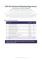
NSG 533 Advanced Pathophysiology Exam 1
Exam Questions and Answers (2026/2027)
Verified Answers By Expert | Complete 125-Question Test Bank
Aligned with NSG 533 Advanced Pathophysiology Course Objectives and Current Pathophysiology Standards
8 Sections | 125 Questions | Cognitive Mix: 30% Recall, 50% Application, 20% Analysis
Independent Study Resource - This exam preparation resource is intended for educational and study purposes to support graduate
nursing students preparing for NSG 533 Advanced Pathophysiology Exam 1. Content is aligned with current pathophysiology
standards, current research in genetics, immunology, and molecular pathophysiology, and graduate-level advanced practice nursing
competencies. Verify all content with current course materials, textbooks, and instructor guidance.
Section and Question Range Mapping
Secti
Topic Questions Count
on
1 Cellular Biology and Adaptation Q1-Q20 20
2 Genetics and Genetic Disorders Q21-Q35 15
3 Inflammation, Immunity, and Infection Q36-Q55 20
4 Fluids, Electrolytes, and Acid-Base Q56-Q72 17
5 Cellular Proliferation and Neoplasia Q73-Q88 16
6 Neurological Pathophysiology Q89-Q102 14
7 Cardiovascular and Respiratory Pathophysiology Q103-Q115 13
8 Endocrine and Renal Pathophysiology Q116-Q125 10
TOTAL 125
Section 1: Section 1: Cellular Biology and Adaptation (Cell Structure, Function,
Adaptation, Injury, & Death)
NSG 533 Advanced Pathophysiology Exam 1 | Page 1
,NSG 533 ADVANCED PATHOPHYSIOLOGY EXAM 1 - 125 QUESTION TEST BANK
Q1: A 65-year-old patient with chronic hypertension has left ventricular hypertrophy (LVH) on
echocardiography. Which cellular adaptation mechanism best explains this finding, and what is the
underlying stimulus?
A. Hyperplasia from increased hormonal stimulation causing an increased number of cells.
B. Hypertrophy from increased workload (pressure overload) causing increased cell size without increased cell
number. [CORRECT]
C. Metaplasia from chronic irritation causing replacement of one cell type with another.
D. Atrophy from decreased workload causing decreased cell size.
Correct Answer: B
Rationale: Hypertrophy is an increase in cell size in response to increased workload, characteristic of cardiac and skeletal
muscle (terminally differentiated cells that cannot undergo hyperplasia). In chronic hypertension, the LV pumps against
elevated afterload, triggering mechanosensors and neurohormonal signaling (angiotensin II, norepinephrine) that activate
signal transduction pathways (MAPK, PI3K/AKT), leading to increased protein synthesis and sarcomere assembly in parallel.
Hyperplasia (Option A) is increased cell number, not applicable to cardiac myocytes. Metaplasia (Option C) is reversible cell
type replacement (e.g., respiratory epithelium in smokers). Atrophy (Option D) is decreased cell size from disuse.
Q2: A 55-year-old male with long-standing gastroesophageal reflux disease (GERD) undergoes upper
endoscopy, which reveals Barrett esophagus (intestinal metaplasia of the distal esophagus). Which
pathophysiologic mechanism best explains this cellular adaptation?
A. Squamous epithelial cells undergo hyperplasia to resist acid damage.
B. Chronic acid exposure reprograms squamous epithelial stem cells to differentiate into intestinal-type
columnar cells (metaplasia) that are more resistant to acid injury. [CORRECT]
C. Esophageal smooth muscle hypertrophies to improve peristalsis.
D. Esophageal cells undergo atrophy due to chronic acid injury.
Correct Answer: B
Rationale: Barrett esophagus is intestinal metaplasia of the distal esophageal squamous epithelium, an adaptive response to
chronic acid exposure. Metaplasia is the reversible replacement of one differentiated cell type with another, often via
reprogramming of stem cells (not transdifferentiation of mature cells). The columnar intestinal-type cells secrete mucin,
conferring resistance to acid. However, this adaptation increases the risk of adenocarcinoma (10-40x baseline). Hyperplasia
(Option A) would not change cell type. Muscle hypertrophy (Option C) and atrophy (Option D) are not the relevant
mechanisms.
Q3: A 70-year-old patient with chronic disuse of the right arm from a stroke exhibits marked muscle wasting.
Which cellular mechanism underlies this adaptation?
A. Hypertrophy from decreased workload.
B. Atrophy from decreased workload, with decreased protein synthesis and increased proteasomal
degradation of contractile proteins. [CORRECT]
C. Apoptosis of motor neurons.
D. Metaplasia of muscle cells.
Correct Answer: B
Rationale: Disuse atrophy occurs when decreased workload reduces mechanical stress signaling, leading to decreased protein
synthesis (via reduced mTOR signaling) and increased ubiquitin-proteasome-mediated degradation of contractile proteins. The
cell number may not decrease immediately; rather, individual myocytes shrink. Apoptosis of motor neurons (Option C) occurs
in denervation atrophy, not pure disuse atrophy. Metaplasia (Option D) involves cell-type change. Hypertrophy (Option A) is
the opposite adaptation.
NSG 533 Advanced Pathophysiology Exam 1 | Page 2
,NSG 533 ADVANCED PATHOPHYSIOLOGY EXAM 1 - 125 QUESTION TEST BANK
Q4: A pathologist examining a cervical biopsy from a 35-year-old woman with HPV infection identifies
dysplastic cells extending through the lower third of the epithelium (CIN I). Which of the following best
describes dysplasia?
A. An irreversible, pre-neoplastic cellular adaptation with normal differentiation.
B. Disordered cellular proliferation with abnormal size, shape, and organization of cells, which may progress
to neoplasia but is potentially reversible if the inciting stimulus is removed. [CORRECT]
C. Replacement of one cell type with another, more resistant type.
D. Increased cell number in response to hormonal stimulation.
Correct Answer: B
Rationale: Dysplasia is disordered cellular proliferation characterized by pleomorphism (variation in size/shape),
hyperchromatic nuclei, increased mitotic figures, and loss of normal polarity. Unlike metaplasia, dysplasia involves deranged
cell growth and is considered pre-neoplastic. Importantly, mild-to-moderate dysplasia is potentially reversible if the stimulus
(e.g., HPV) is removed. CIN I (mild dysplasia) extends through lower third of epithelium. Severe dysplasia (CIN III/carcinoma
in situ) is more likely to progress to invasive carcinoma. Metaplasia (Option C) is cell-type replacement. Hyperplasia (Option
D) is increased cell number.
Q5: A 45-year-old patient who suffered a myocardial infarction has tissue examined microscopically 24 hours
post-infarction. Which cellular finding is characteristic of this early ischemic injury?
A. Coagulative necrosis with preserved tissue architecture and nuclear pyknosis/karyorrhexis. [CORRECT]
B. Liquefactive necrosis with neutrophil infiltration.
C. Caseous necrosis with granulomatous inflammation.
D. Apoptosis with chromatin condensation and apoptotic body formation.
Correct Answer: A
Rationale: Coagulative necrosis is the hallmark of ischemic injury in all tissues except the brain. It results from
hypoxia-induced ATP depletion, failure of membrane Na/K ATPase, cellular swelling, and protein denaturation. The tissue
architecture is preserved (tombstone appearance) because denatured cellular proteins are not immediately degraded. Nuclear
changes (pyknosis, karyorrhexis, karyolysis) occur over 12-24 hours. Neutrophil infiltration begins at 12-24 hours.
Liquefactive necrosis (Option B) occurs in brain ischemia and bacterial abscesses. Caseous necrosis (Option C) is typical of
tuberculosis. Apoptosis (Option D) is programmed cell death without inflammation.
Q6: A 60-year-old patient with type 2 diabetes develops non-enzymatic glycation of proteins via formation of
advanced glycation end-products (AGEs). Which cellular injury mechanism is most directly responsible for
the long-term complications of diabetes (nephropathy, retinopathy, vasculopathy)?
A. Direct oxidative damage to DNA by hyperglycemia.
B. AGEs crosslink collagen and other matrix proteins, activating AGE receptors (RAGE) on endothelial cells,
which triggers NF-kB-mediated inflammation, ROS generation, and fibrosis. [CORRECT]
C. Hyperglycemia-induced apoptosis of beta cells.
D. Direct cytotoxicity to renal podocytes.
Correct Answer: B
Rationale: Advanced glycation end-products (AGEs) form when glucose non-enzymatically reacts with protein amino groups
(Amadori rearrangement). AGEs crosslink long-lived proteins (collagen, elastin), causing vascular stiffening. AGEs also bind
RAGE receptors on endothelial, mesangial, and inflammatory cells, activating NF-kB and triggering chronic inflammation,
oxidative stress, and fibrosis. This drives diabetic nephropathy (mesangial expansion, glomerulosclerosis), retinopathy
(pericyte loss, neovascularization), and macrovascular disease. While oxidative stress (Option A) contributes, the AGE-RAGE
pathway is the central mechanism. Beta cell apoptosis (Option C) occurs in type 1 diabetes. Podocyte toxicity (Option D) is
downstream of glomerular pathology.
NSG 533 Advanced Pathophysiology Exam 1 | Page 3
, NSG 533 ADVANCED PATHOPHYSIOLOGY EXAM 1 - 125 QUESTION TEST BANK
Q7: A 50-year-old male is rescued from a collapsed mine after 24 hours. He develops severe muscle pain, dark
urine, and acute kidney injury. Laboratory findings show markedly elevated creatine kinase (CK) and
potassium. Which cellular injury mechanism is the primary cause of his renal failure?
A. Direct hypoxic injury to renal tubular cells from prolonged ischemia.
B. Rhabdomyolysis with release of myoglobin from necrotic muscle cells, causing intratubular cast formation,
oxidative injury, and vasoconstriction leading to acute tubular necrosis. [CORRECT]
C. Disseminated intravascular coagulation causing renal cortical necrosis.
D. Glomerulonephritis from immune complex deposition.
Correct Answer: B
Rationale: Prolonged compression/immobilization causes crush injury with muscle necrosis (rhabdomyolysis). Released
myoglobin (a heme protein) is filtered by the glomerulus but precipitates in renal tubules (especially with acidic urine),
forming casts that obstruct tubular flow. Myoglobin also induces oxidative injury to tubular epithelial cells and triggers renal
vasoconstriction (via NO scavenging). Hyperkalemia results from muscle cell lysis. Treatment includes aggressive IV fluid
resuscitation, mannitol, and urinary alkalinization. Direct ischemia (Option A) may contribute but is secondary. DIC (Option
C) and glomerulonephritis (Option D) are not the primary mechanisms.
Q8: During a process called autophagy, a cell under starvation conditions degrades its own organelles to
provide substrates for energy production. Which cellular structure is primarily responsible for executing this
process, and what is its physiological significance?
A. The proteasome, which degrades cytosolic proteins tagged with ubiquitin.
B. Lysosomes, which fuse with autophagosomes (double-membrane vesicles containing cytoplasmic cargo) to
degrade damaged organelles and macromolecules during nutrient deprivation. [CORRECT]
C. The Golgi apparatus, which packages proteins for secretion.
D. The endoplasmic reticulum, which synthesizes new proteins.
Correct Answer: B
Rationale: Autophagy (self-eating) is a homeostatic process by which cells degrade damaged organelles (e.g., mitochondria
via mitophagy) and long-lived proteins. Double-membrane autophagosomes engulf cytoplasmic cargo and fuse with
lysosomes, whose acidic hydrolases degrade the contents. Autophagy is upregulated during nutrient starvation (via mTOR
inhibition) to provide amino acids and fatty acids for survival. Dysregulated autophagy is implicated in neurodegeneration
(Alzheimer, Parkinson), cancer, and infection. The proteasome (Option A) degrades short-lived cytosolic proteins tagged with
ubiquitin. The Golgi (Option C) and ER (Option D) handle protein processing and synthesis.
Q9: A 30-year-old patient sustains a spinal cord injury resulting in muscle denervation. Six months later, the
affected muscles show marked atrophy. Which mechanism distinguishes denervation atrophy from disuse
atrophy?
A. Denervation causes hyperplasia, while disuse causes atrophy.
B. Denervation causes apoptosis of muscle fibers, while disuse causes only myofiber shrinkage without cell
death. [CORRECT]
C. Denervation causes hypertrophy, while disuse causes metaplasia.
D. Both are identical at the cellular level.
Correct Answer: B
Rationale: Denervation atrophy is more severe than disuse atrophy because loss of neural trophic signals (e.g., agrin,
neuregulin) triggers apoptotic pathways in myofibers, ultimately leading to cell death and fibro-fatty infiltration. Disuse
atrophy causes myofiber shrinkage (reduced protein synthesis, increased proteolysis) without immediate cell loss; recovery of
activity can restore muscle mass. Denervation atrophy is largely irreversible unless reinnervation occurs (limited by distance
and time). The other options are incorrect regarding the adaptation mechanisms.
NSG 533 Advanced Pathophysiology Exam 1 | Page 4