Exam 1 Study Cardiac
Diastole
§ Ventricular relaxation
§ Filling of ventricles
§ Myocardial tissue perfusion
Systole
§ Ventricular contraction
§ Ejection of blood from ventricles
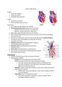
Heart Valves
§ heart needs valves to facilitate one-way flow
§ Atrioventricular (AV) valves between atria & ventricles
o Between R atrium & ventricle is tricuspid valve
o Between L atrium & ventricle is mitral valve
§ When open, valves allow flow from atria into ventricle
§ When ventricles fill & pressure w/in increases, the valves close to prevent retrograde
flow back into atria
§ The valves are connected to muscle in ventricle by fibrous cords, chordae tendineae,
that prevent valves from bulging or turning inside out during
ventricular contraction
§ Semilunar valves between ventricles & their respective
arteries (pulmonary & aortic valves)
§ Pulmonary valve is located between R ventricle &
pulmonary artery
§ Aortic valve is located between L ventricle & aorta
§ Like AV valves, they control flow of blood between ventricles
& pulmonary artery or aorta
Stroke Volume
§ Amount of blood ejected w/ each ventricular contraction
§ Influenced by 3 variables
o Preload: the amount of blood in ventricles @ end of
diastole & amount of stretch of muscle tissue @ end of filling
§ Increased volume produces increased stretch, which produces an
increased contractions; overfilling results in decreased contraction
effectiveness which decreases CO
o Afterload: resistance to flow the ventricle must overcome to open the semilunar
valves & eject its contents
§ Related to BP, vessel lumen diameter, and/or vessel compliance; HTN r/t
increased afterload
o Contractility: force of mechanical contraction
§ Increased w/ sympathetic stimulation or calcium release & can be
decreased w/ hypoxia or acidosis
, Cardiac Output
§ Amount of blood ejected by heart every
minute
§ HR x stroke volume = CO
§ Normal CO in adult is 4-8 L/min
o Body requirements are increased by
exercise or activity & decreased @
rest
Ejection Fraction (EF)
§ Percentage of blood ejected from ventricle w/ each contraction
§ Normal EF is 55-65%
§ <45% indicates some disease process weaking the pumping strength of the heart
§ <30% represents severe decrease in cardiac function & pt @ risk for sudden death
§ Usually measured thru echocardiogram
Echocardiography
§ Transthoracic
o Diagnosis of valve disorders & cardiomyopathy
o Evaluates shape, size, motion of heart
o Measures EF
§ Transesophageal
o Clearer images bc the waves have to pass thru less tissue
o Small transducer is passed thru the mouth into an esophagus
Valve Disorders
§ Abnormality or dysfunction of one of four heart valves
§ Affects the ability of heart to pump & reduces stroke volume
§ Over time, hypertrophy & heart failure can occur
Mitral Valve
§ Valve between L atrium & L ventricle
§ AKA bicuspid (2 flaps/leaflets)
§ In healthy heart, L atrial contraction opens the
mitral valve to allow blood flow into the L ventricle.
Upon completion of ventricular filling, the mitral
valve closes to allow for ventricular contraction to
supply blood to the circulatory system
§ Diseases of mitral valve: regurgitation, stenosis, prolapse
Diastole
§ Ventricular relaxation
§ Filling of ventricles
§ Myocardial tissue perfusion
Systole
§ Ventricular contraction
§ Ejection of blood from ventricles
Heart Valves
§ heart needs valves to facilitate one-way flow
§ Atrioventricular (AV) valves between atria & ventricles
o Between R atrium & ventricle is tricuspid valve
o Between L atrium & ventricle is mitral valve
§ When open, valves allow flow from atria into ventricle
§ When ventricles fill & pressure w/in increases, the valves close to prevent retrograde
flow back into atria
§ The valves are connected to muscle in ventricle by fibrous cords, chordae tendineae,
that prevent valves from bulging or turning inside out during
ventricular contraction
§ Semilunar valves between ventricles & their respective
arteries (pulmonary & aortic valves)
§ Pulmonary valve is located between R ventricle &
pulmonary artery
§ Aortic valve is located between L ventricle & aorta
§ Like AV valves, they control flow of blood between ventricles
& pulmonary artery or aorta
Stroke Volume
§ Amount of blood ejected w/ each ventricular contraction
§ Influenced by 3 variables
o Preload: the amount of blood in ventricles @ end of
diastole & amount of stretch of muscle tissue @ end of filling
§ Increased volume produces increased stretch, which produces an
increased contractions; overfilling results in decreased contraction
effectiveness which decreases CO
o Afterload: resistance to flow the ventricle must overcome to open the semilunar
valves & eject its contents
§ Related to BP, vessel lumen diameter, and/or vessel compliance; HTN r/t
increased afterload
o Contractility: force of mechanical contraction
§ Increased w/ sympathetic stimulation or calcium release & can be
decreased w/ hypoxia or acidosis
, Cardiac Output
§ Amount of blood ejected by heart every
minute
§ HR x stroke volume = CO
§ Normal CO in adult is 4-8 L/min
o Body requirements are increased by
exercise or activity & decreased @
rest
Ejection Fraction (EF)
§ Percentage of blood ejected from ventricle w/ each contraction
§ Normal EF is 55-65%
§ <45% indicates some disease process weaking the pumping strength of the heart
§ <30% represents severe decrease in cardiac function & pt @ risk for sudden death
§ Usually measured thru echocardiogram
Echocardiography
§ Transthoracic
o Diagnosis of valve disorders & cardiomyopathy
o Evaluates shape, size, motion of heart
o Measures EF
§ Transesophageal
o Clearer images bc the waves have to pass thru less tissue
o Small transducer is passed thru the mouth into an esophagus
Valve Disorders
§ Abnormality or dysfunction of one of four heart valves
§ Affects the ability of heart to pump & reduces stroke volume
§ Over time, hypertrophy & heart failure can occur
Mitral Valve
§ Valve between L atrium & L ventricle
§ AKA bicuspid (2 flaps/leaflets)
§ In healthy heart, L atrial contraction opens the
mitral valve to allow blood flow into the L ventricle.
Upon completion of ventricular filling, the mitral
valve closes to allow for ventricular contraction to
supply blood to the circulatory system
§ Diseases of mitral valve: regurgitation, stenosis, prolapse