

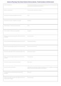
Anatomy & Physiology | Heart, Blood, Endocrine & Nervous Systems - Practice Questions & Verified Answers
What is pericardium? Fluid filled, double walled serous membrane surrounding the heart with parietal and
visceral layers separated by pericardial fluid.
What is endocardium? Tissue lining the heart's four chambers.
What are the 2 superior chambers of the heart? Atria.
What are the 2 inferior chambers of the heart? Ventricles.
Which chamber in the heart receives blood? Atria.
Which chamber in the heart pumps blood? Ventricles.
What structure marks the separation of the atria from the Coronary sulcus.
ventricles?
What structure separates the right ventricle from the left? Interventricular septum.
What structure separates the right atria from the left atria? None of these are correct.
The atria is separated from the ventricles by what valve? Atrioventricular valve.
Which valve lies between the right atrium and the right Tricuspid valve.
ventricle?
Which one is a semi-lunar valve in the heart? Aortic valve.
Describe cardiac muscle tissue. Intercalated discs allow fast electrical transmission, rich in myoglobin and glycogen,
contains many mitochondria, allows synchronization.
Deoxygenated blood enters the heart through what Vena cava.
structure?
What structure takes blood from the right ventricle to the Pulmonary trunk.
lungs to pick up oxygen?
, Anatomy & Physiology | Heart, Blood, Endocrine & Nervous Systems - Practice Questions & Verified Answers
The pulmonary trunk splits into what structure taking the Pulmonary arteries.
blood into the lungs?
What structure returns blood from the lungs to the heart? Pulmonary veins.
Blood is forced into what valve when leaving the left Aortic valve.
ventricle?
What structure takes oxygenated blood from the heart to Aorta.
the rest of the body?
In systemic circulation, what happens to CO2 and O2 in CO2 load and O2 unload.
the blood?
In pulmonary circulation, what happens to CO2 and O2 in Lungs release CO2 and absorb O2.
the blood?
A heart beat is started at what structure? SA Node.
What is considered to be the pacemaker of the heart? SA Node.
What structure sends the electrical impulse to stimulate the AV Node.
ventricles to contract?
What structure sends an electrical impulse to stimulate the SA Node.
atria to contract?
What structure carries the electrical impulse from the AV AV bundle.
node towards the apex of the heart?
What structure fans out from the ends of the AV bundle to Purkinje fibers.
the walls of the ventricle?
What letters are on an EKG waveform? P, Q, R, S, T.
What happens during a P wave? Atrial depolarization.
What happens during the Q, R, S wave? Ventricular depolarization.
, Anatomy & Physiology | Heart, Blood, Endocrine & Nervous Systems - Practice Questions & Verified Answers
What happens during the T wave? Ventricular repolarization.
What is the amount of blood ejected from the ventricle per Stroke volume.
beat?
What chemical doesn't have a positive effect on the heart? Potassium.
What blood vessels carry blood to the heart? Veins.
What blood vessels carry blood away from the heart? Arteries.
What are the 3 types of arteries? Conductor (largest, carotid/pulmonary/aorta), distributor (medium, hepatic/renal),
resistance (smallest, arterioles).
What type of blood vessel has valves that prevent Veins.
backflow of blood?
What are the 5 kinds of venous return? Pressure gradient, gravity, thoracic pump, cardiac suction, skeletal muscle pump.
Blood pressure is measured at which artery? Brachial.
What is a normal blood pressure range? 120/80 or 120/72.
Arter/o artery
Atri/o atrium
Ventricul/o ventricle
Ven/o vein
Tachy rapid
What is pericardium? Fluid filled, double walled serous membrane surrounding the heart with parietal and
visceral layers separated by pericardial fluid.
What is endocardium? Tissue lining the heart's four chambers.
What are the 2 superior chambers of the heart? Atria.
What are the 2 inferior chambers of the heart? Ventricles.
Which chamber in the heart receives blood? Atria.
Which chamber in the heart pumps blood? Ventricles.
What structure marks the separation of the atria from the Coronary sulcus.
ventricles?
What structure separates the right ventricle from the left? Interventricular septum.
What structure separates the right atria from the left atria? None of these are correct.
The atria is separated from the ventricles by what valve? Atrioventricular valve.
Which valve lies between the right atrium and the right Tricuspid valve.
ventricle?
Which one is a semi-lunar valve in the heart? Aortic valve.
Describe cardiac muscle tissue. Intercalated discs allow fast electrical transmission, rich in myoglobin and glycogen,
contains many mitochondria, allows synchronization.
Deoxygenated blood enters the heart through what Vena cava.
structure?
What structure takes blood from the right ventricle to the Pulmonary trunk.
lungs to pick up oxygen?
, Anatomy & Physiology | Heart, Blood, Endocrine & Nervous Systems - Practice Questions & Verified Answers
The pulmonary trunk splits into what structure taking the Pulmonary arteries.
blood into the lungs?
What structure returns blood from the lungs to the heart? Pulmonary veins.
Blood is forced into what valve when leaving the left Aortic valve.
ventricle?
What structure takes oxygenated blood from the heart to Aorta.
the rest of the body?
In systemic circulation, what happens to CO2 and O2 in CO2 load and O2 unload.
the blood?
In pulmonary circulation, what happens to CO2 and O2 in Lungs release CO2 and absorb O2.
the blood?
A heart beat is started at what structure? SA Node.
What is considered to be the pacemaker of the heart? SA Node.
What structure sends the electrical impulse to stimulate the AV Node.
ventricles to contract?
What structure sends an electrical impulse to stimulate the SA Node.
atria to contract?
What structure carries the electrical impulse from the AV AV bundle.
node towards the apex of the heart?
What structure fans out from the ends of the AV bundle to Purkinje fibers.
the walls of the ventricle?
What letters are on an EKG waveform? P, Q, R, S, T.
What happens during a P wave? Atrial depolarization.
What happens during the Q, R, S wave? Ventricular depolarization.
, Anatomy & Physiology | Heart, Blood, Endocrine & Nervous Systems - Practice Questions & Verified Answers
What happens during the T wave? Ventricular repolarization.
What is the amount of blood ejected from the ventricle per Stroke volume.
beat?
What chemical doesn't have a positive effect on the heart? Potassium.
What blood vessels carry blood to the heart? Veins.
What blood vessels carry blood away from the heart? Arteries.
What are the 3 types of arteries? Conductor (largest, carotid/pulmonary/aorta), distributor (medium, hepatic/renal),
resistance (smallest, arterioles).
What type of blood vessel has valves that prevent Veins.
backflow of blood?
What are the 5 kinds of venous return? Pressure gradient, gravity, thoracic pump, cardiac suction, skeletal muscle pump.
Blood pressure is measured at which artery? Brachial.
What is a normal blood pressure range? 120/80 or 120/72.
Arter/o artery
Atri/o atrium
Ventricul/o ventricle
Ven/o vein
Tachy rapid


