
AAPC ACTUAL SCRIPT WITH 100
PERCENT CORRECT ANSWERS
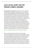
◉ PREOPERATIVE DIAGNOSIS: Medial meniscus tear, right knee
POSTOPERATIVE DIAGNOSIS: Medial meniscus tear, extensive
synovitis with an impingement medial synovial plica, right knee
TITLE OF PROCEDURE: Diagnostic operative arthroscopy, partial
medial meniscectomy and synovectomy, right knee The patent
was brought to the operating room, placed in the supine position
after which he underwent general anesthesia. The right knee was
then prepped and draped in the usual sterile fashion. The
arthroscope was introduced through an anterolateral portal,
interim portal created anteromedially. The suprapatellar pouch
was inspected. The findings on the patella and the femoral groove
were as noted above. An intra-articular shaver was introduced to
debride the loose fibrillated articular cartilage from the medial
patellar facet. The hypertrophic synovial scarring between the
patella and the femoral groove was debrided. The hypertrophic
Answer: C. 29881-RT, M23.221, M65.861, M94.261, M22.41
◉ A 61-year-old gentleman with a history of a fall while
intoxicated suffered a blow to the forehead and imaging revealed
a posteriorly displaced odontoid fracture. The patient was taken
into the Operating Room, and placed supine on the operating
room table. Under mild sedation, the patient was placed in
Gardner-Wells tongs and gentle axial traction under fluoroscopy
was performed to gently try to reduce the fracture. It did reduce
partially without any change in the neurologic examination. More
manipulation would be necessary and it was decided to intubate
,and use fiberoptic technique. The anterior neck was prepped and
draped and an incision was made in a skin crease overlying the
C4-C5 area. Using hand-held retractors, the ventral aspect of the
spine was identified and the C2-C3 disk space was identified using
lateral fluoroscopy. Using some pressure upon the ventral aspect
of the C2 body, we were able to achieve a s
Answer: D. 22318
◉ The patient is a 51-year-old gentleman who has end-stage renal
disease. He was in the OR yesterday for a revision of his AV graft.
The next day the patient had complications of the graft failing. The
patient was back to the operating room where an open
thrombectomy was performed on both sides getting good back
bleeding, good inflow. Select the appropriate code for performing
the procedure in a post-operative period:
A. 36831-76
B. 36831
C. 36831-78
D. 36831-58
Answer: C. 36831-78
◉ The patient is a 77-year-old white female who has been having
right temporal pain and headaches with some visual changes and
has a sed rate of 51. She is scheduled for a temporal artery biopsy
to rule out temporal arteritis. A Doppler probe was used to isolate
the temporal artery and using a marking pen the path of the
artery was drawn. Lidocaine 1% was used to infiltrate the skin,
and using a 15 blade scalpel the skin was opened in the
, preauricular area and dissected down to the subcutaneous tissue
where the temporal artery was identified in its bed. It was a
medium size artery and we dissected it out for a length of
approximately 4 cm with some branches. The ends were ligated
with 4-0 Vicryl, and the artery was removed from its bed and sent
to Pathology as specimen. What CPT® code is reported?
A. 37609
B. 37605
C. 36625
D. 37799
Answer: A. 37609
◉ A 50-year-old female has recurrent lymphoma in the axilla.
Ultrasound was used to localize the lymph node in question for
needle guidance. An 11 blade scalpel was used to perform a small
dermatotomy. An 18 x 10 cm Biopence needle was advanced
through the dermatotomy to the periphery of the lymph node. A
total of 4 biopsy specimens were obtained. Two specimens were
placed an RPMI and 2 were placed in formalin and sent to
laboratory. The correct CPT® code(s) is (are):
A. 10005
B. 38500, 77002-26
C. 38505, 76942-26
D. 38525, 76942-26
Answer: C. 38505, 76942-26
PERCENT CORRECT ANSWERS
◉ PREOPERATIVE DIAGNOSIS: Medial meniscus tear, right knee
POSTOPERATIVE DIAGNOSIS: Medial meniscus tear, extensive
synovitis with an impingement medial synovial plica, right knee
TITLE OF PROCEDURE: Diagnostic operative arthroscopy, partial
medial meniscectomy and synovectomy, right knee The patent
was brought to the operating room, placed in the supine position
after which he underwent general anesthesia. The right knee was
then prepped and draped in the usual sterile fashion. The
arthroscope was introduced through an anterolateral portal,
interim portal created anteromedially. The suprapatellar pouch
was inspected. The findings on the patella and the femoral groove
were as noted above. An intra-articular shaver was introduced to
debride the loose fibrillated articular cartilage from the medial
patellar facet. The hypertrophic synovial scarring between the
patella and the femoral groove was debrided. The hypertrophic
Answer: C. 29881-RT, M23.221, M65.861, M94.261, M22.41
◉ A 61-year-old gentleman with a history of a fall while
intoxicated suffered a blow to the forehead and imaging revealed
a posteriorly displaced odontoid fracture. The patient was taken
into the Operating Room, and placed supine on the operating
room table. Under mild sedation, the patient was placed in
Gardner-Wells tongs and gentle axial traction under fluoroscopy
was performed to gently try to reduce the fracture. It did reduce
partially without any change in the neurologic examination. More
manipulation would be necessary and it was decided to intubate
,and use fiberoptic technique. The anterior neck was prepped and
draped and an incision was made in a skin crease overlying the
C4-C5 area. Using hand-held retractors, the ventral aspect of the
spine was identified and the C2-C3 disk space was identified using
lateral fluoroscopy. Using some pressure upon the ventral aspect
of the C2 body, we were able to achieve a s
Answer: D. 22318
◉ The patient is a 51-year-old gentleman who has end-stage renal
disease. He was in the OR yesterday for a revision of his AV graft.
The next day the patient had complications of the graft failing. The
patient was back to the operating room where an open
thrombectomy was performed on both sides getting good back
bleeding, good inflow. Select the appropriate code for performing
the procedure in a post-operative period:
A. 36831-76
B. 36831
C. 36831-78
D. 36831-58
Answer: C. 36831-78
◉ The patient is a 77-year-old white female who has been having
right temporal pain and headaches with some visual changes and
has a sed rate of 51. She is scheduled for a temporal artery biopsy
to rule out temporal arteritis. A Doppler probe was used to isolate
the temporal artery and using a marking pen the path of the
artery was drawn. Lidocaine 1% was used to infiltrate the skin,
and using a 15 blade scalpel the skin was opened in the
, preauricular area and dissected down to the subcutaneous tissue
where the temporal artery was identified in its bed. It was a
medium size artery and we dissected it out for a length of
approximately 4 cm with some branches. The ends were ligated
with 4-0 Vicryl, and the artery was removed from its bed and sent
to Pathology as specimen. What CPT® code is reported?
A. 37609
B. 37605
C. 36625
D. 37799
Answer: A. 37609
◉ A 50-year-old female has recurrent lymphoma in the axilla.
Ultrasound was used to localize the lymph node in question for
needle guidance. An 11 blade scalpel was used to perform a small
dermatotomy. An 18 x 10 cm Biopence needle was advanced
through the dermatotomy to the periphery of the lymph node. A
total of 4 biopsy specimens were obtained. Two specimens were
placed an RPMI and 2 were placed in formalin and sent to
laboratory. The correct CPT® code(s) is (are):
A. 10005
B. 38500, 77002-26
C. 38505, 76942-26
D. 38525, 76942-26
Answer: C. 38505, 76942-26


