
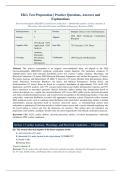
EKG Test Preparation | Practice Questions, Answers and
Explanations
Electrocardiography (EKG/ECG) Technician Certification — Rhythm Recognition, Cardiac Anatomy &
Physiology, Electrode Placement, and Medical Emergency Response (2026)
Total Questions: 75 Format: Multiple Choice (A-D) with Rationales
2026 EKG Technician Certification
Sections: 5 Aligned:
Blueprint
30% Recall / 50%
Cognitive Mix: Application / 20% Item Mix: 75% Scenario / 25% Direct
Analysis
Anatomy,
Electrodes,
Coverage: Waveforms, Verification: 100% Keyed with Rationales
Rhythms,
Emergencies
Abstract. This practice examination is an original, non-confidential study aid aligned to the 2026
electrocardiography (EKG/ECG) technician certification content blueprint. The instrument comprises 75
multiple-choice items with rationales distributed across five sections: Cardiac Anatomy, Physiology, and
Electrical Conduction (15 items); EKG Electrode Placement, Equipment, and Artifact Recognition (15 items);
Waveform Analysis and Measurement (P, QRS, T, Intervals) (15 items); Rhythm Strip Interpretation (Sinus,
Atrial, Junctional, Ventricular Rhythms) (18 items); and Medical Emergencies, Patient Safety, and
Professionalism (12 items). Items are keyed to a cognitive distribution of approximately 30% recall, 50%
application, and 20% analysis, with 75% scenario-based clinical and rhythm interpretation vignettes and 25%
direct anatomical or procedural questions. Special inclusions address rhythm strip interpretation based on
written descriptions (e.g., regular rate, presence/absence of P waves, QRS width), precise electrode placement
and artifact troubleshooting/correction, and scenario-based recognition of life-threatening rhythms (ventricular
tachycardia, ventricular fibrillation, asystole) with appropriate immediate actions. Distractors reflect common
EKG technician errors, such as confusing atrial fibrillation with atrial flutter, misidentifying artifact as rhythm
abnormalities, placing precordial leads in incorrect intercostal spaces, or misinterpreting normal sinus
arrhythmia as pathological. Each item includes a verified correct answer and a concise rationale explaining why
the keyed option is correct and why the distractors are incorrect. This artifact does not reproduce any
proprietary exam content; candidates should verify all current requirements with official certification materials.
Keywords: EKG; ECG; cardiac rhythm; electrode placement; artifact; waveform interpretation; ventricular
fibrillation; CPR; cardiac conduction
Section 1: Cardiac Anatomy, Physiology, and Electrical Conduction — 15 Questions
Q1: The normal electrical impulse of the heart originates in the:
A. Atrioventricular (AV) node
B. Sinoatrial (SA) node, located in the right atrium *[CORRECT]*
C. Bundle of His
D. Purkinje fibers
Correct Answer: B
Rationale: The SA node in the right atrium is the heart's natural pacemaker, initiating 60-100 impulses per minute;
the AV node, bundle of His, and Purkinje fibers conduct but do not normally initiate.
,Q2: Which structure delays the electrical impulse briefly to allow the atria to complete contraction
before ventricular depolarization?
A. The SA node
B. The bundle of His
C. The atrioventricular (AV) node *[CORRECT]*
D. The Purkinje fibers
Correct Answer: C
Rationale: The AV node delays the impulse ~0.1 second allowing atrial systole to complete before ventricular
activation; the SA node, bundle of His, and Purkinje fibers do not provide this delay.
Q3: The normal intrinsic rate of the SA node is:
A. 40-60 beats per minute
B. 20-40 beats per minute
C. 100-150 beats per minute
D. 60-100 beats per minute *[CORRECT]*
Correct Answer: D
Rationale: The SA node fires at 60-100 bpm; the AV junction fires 40-60, Purkinje/ventricular 20-40, and 100-150
would be tachycardic.
Q4: The Purkinje fibers are responsible for:
A. Rapidly distributing the impulse through the ventricles to coordinate contraction *[CORRECT]*
B. Generating the primary pacemaker impulse
C. Delaying the impulse at the AV node
D. Pumping blood through the aorta
Correct Answer: A
Rationale: Purkinje fibers rapidly distribute the impulse throughout the ventricles for coordinated contraction; they
do not generate primary pacemaker impulses, delay at the AV node, or pump blood.
Q5: Depolarization of the atria produces which waveform on the EKG?
A. The QRS complex
B. The P wave *[CORRECT]*
C. The T wave
D. The PR segment
Correct Answer: B
Rationale: Atrial depolarization produces the P wave; ventricular depolarization produces the QRS, ventricular
repolarization the T wave, and the PR segment is the delay.
Q6: The QRS complex on an EKG represents:
A. Atrial repolarization
B. Ventricular repolarization
C. Ventricular depolarization *[CORRECT]*
D. Atrial depolarization
Correct Answer: C
Rationale: The QRS represents ventricular depolarization; atrial repolarization is hidden in the QRS, ventricular
repolarization is the T wave, and atrial depolarization is the P wave.
Q7: The T wave represents:
A. Atrial depolarization
B. Ventricular depolarization
, C. Atrial repolarization
D. Ventricular repolarization *[CORRECT]*
Correct Answer: D
Rationale: The T wave represents ventricular repolarization; atrial depolarization is the P wave, ventricular
depolarization is the QRS, and atrial repolarization is hidden.
Q8: The normal heart has how many chambers?
A. Four: two atria and two ventricles *[CORRECT]*
B. Two
C. Three
D. Six
Correct Answer: A
Rationale: The heart has four chambers (right/left atria, right/left ventricles); two, three, and six are incorrect.
Q9: Which valve prevents backflow from the left ventricle to the left atrium?
A. The tricuspid valve
B. The mitral (bicuspid) valve *[CORRECT]*
C. The aortic valve
D. The pulmonic valve
Correct Answer: B
Rationale: The mitral (bicuspid) valve is between the left atrium and left ventricle; the tricuspid is on the right, the
aortic and pulmonic are semilunar outflow valves.
Q10: The right atrium receives deoxygenated blood from:
A. The pulmonary veins
B. The aorta
C. The superior and inferior vena cavae *[CORRECT]*
D. The coronary sinus only
Correct Answer: C
Rationale: The right atrium receives deoxygenated blood from the superior and inferior vena cavae; the pulmonary
veins return oxygenated blood to the left atrium, the aorta carries oxygenated blood out, and the coronary sinus is a
minor contributor.
Q11: The left ventricle pumps blood to:
A. The pulmonary circulation via the pulmonary artery
B. The right atrium
C. The lungs only
D. The systemic circulation via the aorta *[CORRECT]*
Correct Answer: D
Rationale: The left ventricle pumps oxygenated blood to the systemic circulation via the aorta; the right ventricle
pumps to the pulmonary circulation, and the right atrium/lungs are not left-ventricular destinations.
Q12: The bundle of His is located:
A. In the interventricular septum, connecting the AV node to the bundle branches *[CORRECT]*
B. In the right atrium
C. In the pulmonary artery
D. In the aortic arch
Correct Answer: A
Explanations
Electrocardiography (EKG/ECG) Technician Certification — Rhythm Recognition, Cardiac Anatomy &
Physiology, Electrode Placement, and Medical Emergency Response (2026)
Total Questions: 75 Format: Multiple Choice (A-D) with Rationales
2026 EKG Technician Certification
Sections: 5 Aligned:
Blueprint
30% Recall / 50%
Cognitive Mix: Application / 20% Item Mix: 75% Scenario / 25% Direct
Analysis
Anatomy,
Electrodes,
Coverage: Waveforms, Verification: 100% Keyed with Rationales
Rhythms,
Emergencies
Abstract. This practice examination is an original, non-confidential study aid aligned to the 2026
electrocardiography (EKG/ECG) technician certification content blueprint. The instrument comprises 75
multiple-choice items with rationales distributed across five sections: Cardiac Anatomy, Physiology, and
Electrical Conduction (15 items); EKG Electrode Placement, Equipment, and Artifact Recognition (15 items);
Waveform Analysis and Measurement (P, QRS, T, Intervals) (15 items); Rhythm Strip Interpretation (Sinus,
Atrial, Junctional, Ventricular Rhythms) (18 items); and Medical Emergencies, Patient Safety, and
Professionalism (12 items). Items are keyed to a cognitive distribution of approximately 30% recall, 50%
application, and 20% analysis, with 75% scenario-based clinical and rhythm interpretation vignettes and 25%
direct anatomical or procedural questions. Special inclusions address rhythm strip interpretation based on
written descriptions (e.g., regular rate, presence/absence of P waves, QRS width), precise electrode placement
and artifact troubleshooting/correction, and scenario-based recognition of life-threatening rhythms (ventricular
tachycardia, ventricular fibrillation, asystole) with appropriate immediate actions. Distractors reflect common
EKG technician errors, such as confusing atrial fibrillation with atrial flutter, misidentifying artifact as rhythm
abnormalities, placing precordial leads in incorrect intercostal spaces, or misinterpreting normal sinus
arrhythmia as pathological. Each item includes a verified correct answer and a concise rationale explaining why
the keyed option is correct and why the distractors are incorrect. This artifact does not reproduce any
proprietary exam content; candidates should verify all current requirements with official certification materials.
Keywords: EKG; ECG; cardiac rhythm; electrode placement; artifact; waveform interpretation; ventricular
fibrillation; CPR; cardiac conduction
Section 1: Cardiac Anatomy, Physiology, and Electrical Conduction — 15 Questions
Q1: The normal electrical impulse of the heart originates in the:
A. Atrioventricular (AV) node
B. Sinoatrial (SA) node, located in the right atrium *[CORRECT]*
C. Bundle of His
D. Purkinje fibers
Correct Answer: B
Rationale: The SA node in the right atrium is the heart's natural pacemaker, initiating 60-100 impulses per minute;
the AV node, bundle of His, and Purkinje fibers conduct but do not normally initiate.
,Q2: Which structure delays the electrical impulse briefly to allow the atria to complete contraction
before ventricular depolarization?
A. The SA node
B. The bundle of His
C. The atrioventricular (AV) node *[CORRECT]*
D. The Purkinje fibers
Correct Answer: C
Rationale: The AV node delays the impulse ~0.1 second allowing atrial systole to complete before ventricular
activation; the SA node, bundle of His, and Purkinje fibers do not provide this delay.
Q3: The normal intrinsic rate of the SA node is:
A. 40-60 beats per minute
B. 20-40 beats per minute
C. 100-150 beats per minute
D. 60-100 beats per minute *[CORRECT]*
Correct Answer: D
Rationale: The SA node fires at 60-100 bpm; the AV junction fires 40-60, Purkinje/ventricular 20-40, and 100-150
would be tachycardic.
Q4: The Purkinje fibers are responsible for:
A. Rapidly distributing the impulse through the ventricles to coordinate contraction *[CORRECT]*
B. Generating the primary pacemaker impulse
C. Delaying the impulse at the AV node
D. Pumping blood through the aorta
Correct Answer: A
Rationale: Purkinje fibers rapidly distribute the impulse throughout the ventricles for coordinated contraction; they
do not generate primary pacemaker impulses, delay at the AV node, or pump blood.
Q5: Depolarization of the atria produces which waveform on the EKG?
A. The QRS complex
B. The P wave *[CORRECT]*
C. The T wave
D. The PR segment
Correct Answer: B
Rationale: Atrial depolarization produces the P wave; ventricular depolarization produces the QRS, ventricular
repolarization the T wave, and the PR segment is the delay.
Q6: The QRS complex on an EKG represents:
A. Atrial repolarization
B. Ventricular repolarization
C. Ventricular depolarization *[CORRECT]*
D. Atrial depolarization
Correct Answer: C
Rationale: The QRS represents ventricular depolarization; atrial repolarization is hidden in the QRS, ventricular
repolarization is the T wave, and atrial depolarization is the P wave.
Q7: The T wave represents:
A. Atrial depolarization
B. Ventricular depolarization
, C. Atrial repolarization
D. Ventricular repolarization *[CORRECT]*
Correct Answer: D
Rationale: The T wave represents ventricular repolarization; atrial depolarization is the P wave, ventricular
depolarization is the QRS, and atrial repolarization is hidden.
Q8: The normal heart has how many chambers?
A. Four: two atria and two ventricles *[CORRECT]*
B. Two
C. Three
D. Six
Correct Answer: A
Rationale: The heart has four chambers (right/left atria, right/left ventricles); two, three, and six are incorrect.
Q9: Which valve prevents backflow from the left ventricle to the left atrium?
A. The tricuspid valve
B. The mitral (bicuspid) valve *[CORRECT]*
C. The aortic valve
D. The pulmonic valve
Correct Answer: B
Rationale: The mitral (bicuspid) valve is between the left atrium and left ventricle; the tricuspid is on the right, the
aortic and pulmonic are semilunar outflow valves.
Q10: The right atrium receives deoxygenated blood from:
A. The pulmonary veins
B. The aorta
C. The superior and inferior vena cavae *[CORRECT]*
D. The coronary sinus only
Correct Answer: C
Rationale: The right atrium receives deoxygenated blood from the superior and inferior vena cavae; the pulmonary
veins return oxygenated blood to the left atrium, the aorta carries oxygenated blood out, and the coronary sinus is a
minor contributor.
Q11: The left ventricle pumps blood to:
A. The pulmonary circulation via the pulmonary artery
B. The right atrium
C. The lungs only
D. The systemic circulation via the aorta *[CORRECT]*
Correct Answer: D
Rationale: The left ventricle pumps oxygenated blood to the systemic circulation via the aorta; the right ventricle
pumps to the pulmonary circulation, and the right atrium/lungs are not left-ventricular destinations.
Q12: The bundle of His is located:
A. In the interventricular septum, connecting the AV node to the bundle branches *[CORRECT]*
B. In the right atrium
C. In the pulmonary artery
D. In the aortic arch
Correct Answer: A


