
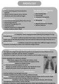
RADIOLOGY
PACS
Picture Archiving and Communication Three Modes of Image Production
Systems →Transmission of energy
System used to acquire, store, retrieve, Plain Radiography
distribute, and view medical images Fluoroscopy
Computed Tomography
Radiologic Modalities →Reflection of energy
Conventional Radiography/Plain Films (X-ray) Ultrasound
Fluoroscopy (Fluoro) →Emission of energy
Computed Tomography (CT) Magnetic Resonance Imaging
Ultrasound (US) Nuclear Medicine
Magnetic Resonance Imaging (MRI)
Nuclear Medicine (Nucl. Med)
Transmission of Energy
Plain radiographs – a “snapshot”, static image generated following the passage of X-rays
through the pt
Fluoroscopy – a “movie”, medical procedure that makes a real-time video of the movements
inside a part of the body by passing X-rays and contrast material through the body over a
period of time
Computed Tomography – combines a series of X-rays taken from different angles and uses
computer processing to create cross-sectional images (slices)
Radiograph: a beam of high-energy photons is passed through the body
The higher the weight through which the photons are passed, the more likely it is to be
attenuated or “blocked” and the “denser” it appears
The denser an object is, the more X-rays it absorbs, and the whiter it appears
→ 5 basic densities from whitest to darkest:
Metal- Most dense, appears the whitest (ex: bullets, coins)
Calcium- Contained within bones, is dense and appears white
Fluid or soft tissue- Appears as shades of gray.
Fluid and soft tissues appear the same
Fat- Appears as shades of gray but darker than fluid/soft tissues
Air- Absorbs the least amount and appears the darkest
Ionizing Radiation
Ionizing radiation can damage living cells
Imaging modalities that produce ionizing radiation include X-ray, CT, Fluoro, & Nucl. Med.
Benefits= X-rays/CT have the ability to diagnose disease, Detect lesions sooner, Stage
disease more accurately, Direct therapy, & Monitor respones
Disadvantages= Radiation sickness, Radiation-induced thyroid disease, Skin damage,
Teratogenesis, Birth defects, Cancer, Sterility
Radiation Protection= Avoid repeated exposures, Wear lead shielding, Avoid in pregnancy
, Computed Tomography: uses a rotating X-ray beam to take a series of X-rays and uses
computer processing to create cross-sectional images or “slices”
These slices are called tomographic images and can give a clinician more detailed
information than conventional X-rays
Images can also be formatted in different planes: Coronal, Axial, Sagittal
Post-processing computers also produce “windows” to view specific structures
CT is moderately expensive and uses ionizing radiation but has many uses
Reflection Imaging- Ultrasound: utilizes high-frequency sound waves to create an image of
the inside of the body
A transducer/ probe transmits sound waves and record the waves that reflect/echo back
The shape & intensity of the echoes depend on how the sound waves interact with
different tissues
For example, most waves pass through a fluid-filled cyst and send back very few or faint
echoes, which look black on the display screen
On the other hand, waves will bounce off a solid tumor, creating a pattern of echoes that
the computer will interpret as a lighter-colored image
US has many uses, It is relatively inexpensive compared to CT and MRI, widely available,
portable, and safe (utilizes no ionizing radiation)
Emission Imaging
Magnetic Resonance Imaging: uses a magnetic field and computer-generated radio waves to
create detailed images of organs and tissues
Provides superior soft tissue differentiation; does NOT utilize ionizing radiation
The strong magnetic field created by the MRI scanner causes the H+ atoms in the body to
align in the same direction; Radiofrequency waves are sent from the MRI machine,
temporarily disturbing the alignment of hydrogen nuclei.
When the radio waves are turned off, the hydrogen nuclei return to their original alignment
and release signals that are converted into an image by a computer.
MRI has many uses: widely used in neurologic imaging and is particularly sensitive in
imaging the brain, spinal cord, and nerves
MRA (magnetic resonance angiography): uses an IV injection of contrast into the blood
vessels and MRI scanning to visualize blood vessels
MRI disadvantages: Relatively expensive, Time-consuming
MRI limitations: Inferior to CT in detecting acute hemorrhage; Inferior to CT in detection of
bone fractures; Metallic implants and devices require careful screening
Nuclear Medicine: specialized area of radiology that uses very small amounts of radioactive
materials to examine organ function and structure
Adds a dimension to medical imaging, by adding the ability to evaluate physiology
Does utilize ionizing radiation; It is a painless procedure and relatively safe
Various body organs have a specific affinity for, or absorption of, different biologically
active chemicals
Common uses: Bone scans, Cardiac scans , Thyroid scans , HIDA scans PET scan
Contrast agents play a crucial role in imaging by improving anatomical visibility of bodily
structures or fluids
, Types of Contrast
Most common routes of administration are: Intravenous (IV), Oral (PO), Rectal
Iodinated contrast media Iodine is the major component. Ex: gastrografin
Non-iodinated contrast media Does not contain any iodine. Ex: barium
Gadolinium-based Most are water soluble, used in MRIs
Negative contrast agents Used to make tissues and structures appear darker which creates
contrast between the agent and surrounding tissues (air, oxygen, carbon dioxide, and nitrous
oxide)
Iodine (X-rays & CTs)- enhances blood vessels or organs
Ex: CT abdomen/pelvis with contrast
Also utilized in fluoroscopy, angiography and venography
Barium (X-rays & CTs)
Used most often in the GI tract
Ex: barium swallow, barium enema, etc
Discerns structural features, including pathologic lesions
Excellent for discerning large masses and strictures
Gadolinium (MRIs)
Improves the diagnostic accuracy of an MRI by improving the visibility of inflammation,
tumors, and blood vessels
Administered only via IV
Risks of Contrast Agents
Gadolinium reactions are rare and typically mild
Iodinated reactions are more frequent and more severe
Contrast-induced kidney injury can also occur
, POCUS
Impedance: resistance of sound waves through fluids
→ Different Modes: A, B, M, Doppler, Duplex
→ Different Probes:
Curvilinear probe: low frequency, wide footprint, deep
structures
Linear probe: high frequency, linear footprint, shallow
structures
Phased Array Probe: low frequency, narrow footprint,
deep structures
→ Advantages of US:
No ionizing radiation, no long term side effects, “real
time” images, and produces little or no discomfort
→ Disadvantages of US:
Difficult penetrating through bone, gas-filled
structures reduce urs utility, obese patient may be
difficult to penetrate, dependent on the skills of the
operator scanning
→ Nomenclature:
Anechoic: black
Hyperchoic (brighter) compared to surrounding tissue
Hypoechoic (darker)
Isochoic (similar)
→ FAST Exam
Focused Assessment with Sonography in Trauma
Mostly for abdominal & thoracic trauma
Image Artifact
Mirror Image →
Refraction →
Acoustic Shadowing →
Comet Tail →
PACS
Picture Archiving and Communication Three Modes of Image Production
Systems →Transmission of energy
System used to acquire, store, retrieve, Plain Radiography
distribute, and view medical images Fluoroscopy
Computed Tomography
Radiologic Modalities →Reflection of energy
Conventional Radiography/Plain Films (X-ray) Ultrasound
Fluoroscopy (Fluoro) →Emission of energy
Computed Tomography (CT) Magnetic Resonance Imaging
Ultrasound (US) Nuclear Medicine
Magnetic Resonance Imaging (MRI)
Nuclear Medicine (Nucl. Med)
Transmission of Energy
Plain radiographs – a “snapshot”, static image generated following the passage of X-rays
through the pt
Fluoroscopy – a “movie”, medical procedure that makes a real-time video of the movements
inside a part of the body by passing X-rays and contrast material through the body over a
period of time
Computed Tomography – combines a series of X-rays taken from different angles and uses
computer processing to create cross-sectional images (slices)
Radiograph: a beam of high-energy photons is passed through the body
The higher the weight through which the photons are passed, the more likely it is to be
attenuated or “blocked” and the “denser” it appears
The denser an object is, the more X-rays it absorbs, and the whiter it appears
→ 5 basic densities from whitest to darkest:
Metal- Most dense, appears the whitest (ex: bullets, coins)
Calcium- Contained within bones, is dense and appears white
Fluid or soft tissue- Appears as shades of gray.
Fluid and soft tissues appear the same
Fat- Appears as shades of gray but darker than fluid/soft tissues
Air- Absorbs the least amount and appears the darkest
Ionizing Radiation
Ionizing radiation can damage living cells
Imaging modalities that produce ionizing radiation include X-ray, CT, Fluoro, & Nucl. Med.
Benefits= X-rays/CT have the ability to diagnose disease, Detect lesions sooner, Stage
disease more accurately, Direct therapy, & Monitor respones
Disadvantages= Radiation sickness, Radiation-induced thyroid disease, Skin damage,
Teratogenesis, Birth defects, Cancer, Sterility
Radiation Protection= Avoid repeated exposures, Wear lead shielding, Avoid in pregnancy
, Computed Tomography: uses a rotating X-ray beam to take a series of X-rays and uses
computer processing to create cross-sectional images or “slices”
These slices are called tomographic images and can give a clinician more detailed
information than conventional X-rays
Images can also be formatted in different planes: Coronal, Axial, Sagittal
Post-processing computers also produce “windows” to view specific structures
CT is moderately expensive and uses ionizing radiation but has many uses
Reflection Imaging- Ultrasound: utilizes high-frequency sound waves to create an image of
the inside of the body
A transducer/ probe transmits sound waves and record the waves that reflect/echo back
The shape & intensity of the echoes depend on how the sound waves interact with
different tissues
For example, most waves pass through a fluid-filled cyst and send back very few or faint
echoes, which look black on the display screen
On the other hand, waves will bounce off a solid tumor, creating a pattern of echoes that
the computer will interpret as a lighter-colored image
US has many uses, It is relatively inexpensive compared to CT and MRI, widely available,
portable, and safe (utilizes no ionizing radiation)
Emission Imaging
Magnetic Resonance Imaging: uses a magnetic field and computer-generated radio waves to
create detailed images of organs and tissues
Provides superior soft tissue differentiation; does NOT utilize ionizing radiation
The strong magnetic field created by the MRI scanner causes the H+ atoms in the body to
align in the same direction; Radiofrequency waves are sent from the MRI machine,
temporarily disturbing the alignment of hydrogen nuclei.
When the radio waves are turned off, the hydrogen nuclei return to their original alignment
and release signals that are converted into an image by a computer.
MRI has many uses: widely used in neurologic imaging and is particularly sensitive in
imaging the brain, spinal cord, and nerves
MRA (magnetic resonance angiography): uses an IV injection of contrast into the blood
vessels and MRI scanning to visualize blood vessels
MRI disadvantages: Relatively expensive, Time-consuming
MRI limitations: Inferior to CT in detecting acute hemorrhage; Inferior to CT in detection of
bone fractures; Metallic implants and devices require careful screening
Nuclear Medicine: specialized area of radiology that uses very small amounts of radioactive
materials to examine organ function and structure
Adds a dimension to medical imaging, by adding the ability to evaluate physiology
Does utilize ionizing radiation; It is a painless procedure and relatively safe
Various body organs have a specific affinity for, or absorption of, different biologically
active chemicals
Common uses: Bone scans, Cardiac scans , Thyroid scans , HIDA scans PET scan
Contrast agents play a crucial role in imaging by improving anatomical visibility of bodily
structures or fluids
, Types of Contrast
Most common routes of administration are: Intravenous (IV), Oral (PO), Rectal
Iodinated contrast media Iodine is the major component. Ex: gastrografin
Non-iodinated contrast media Does not contain any iodine. Ex: barium
Gadolinium-based Most are water soluble, used in MRIs
Negative contrast agents Used to make tissues and structures appear darker which creates
contrast between the agent and surrounding tissues (air, oxygen, carbon dioxide, and nitrous
oxide)
Iodine (X-rays & CTs)- enhances blood vessels or organs
Ex: CT abdomen/pelvis with contrast
Also utilized in fluoroscopy, angiography and venography
Barium (X-rays & CTs)
Used most often in the GI tract
Ex: barium swallow, barium enema, etc
Discerns structural features, including pathologic lesions
Excellent for discerning large masses and strictures
Gadolinium (MRIs)
Improves the diagnostic accuracy of an MRI by improving the visibility of inflammation,
tumors, and blood vessels
Administered only via IV
Risks of Contrast Agents
Gadolinium reactions are rare and typically mild
Iodinated reactions are more frequent and more severe
Contrast-induced kidney injury can also occur
, POCUS
Impedance: resistance of sound waves through fluids
→ Different Modes: A, B, M, Doppler, Duplex
→ Different Probes:
Curvilinear probe: low frequency, wide footprint, deep
structures
Linear probe: high frequency, linear footprint, shallow
structures
Phased Array Probe: low frequency, narrow footprint,
deep structures
→ Advantages of US:
No ionizing radiation, no long term side effects, “real
time” images, and produces little or no discomfort
→ Disadvantages of US:
Difficult penetrating through bone, gas-filled
structures reduce urs utility, obese patient may be
difficult to penetrate, dependent on the skills of the
operator scanning
→ Nomenclature:
Anechoic: black
Hyperchoic (brighter) compared to surrounding tissue
Hypoechoic (darker)
Isochoic (similar)
→ FAST Exam
Focused Assessment with Sonography in Trauma
Mostly for abdominal & thoracic trauma
Image Artifact
Mirror Image →
Refraction →
Acoustic Shadowing →
Comet Tail →


