
HIGH-YIELD A&P | NERVOUS TISSUE & ACTION POTENTIALS OpenStax 2e | HAPS | TEAS / HESI
MODULE 01 | CHAPTER 12
NERVOUS TISSUE & ACTION POTENTIALS
High-Yield Study Guide | OpenStax Anatomy and Physiology 2e | HAPS-Aligned
CORE CONCEPT OVERVIEW
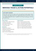
Nervous tissue is built from two cell types: neurons, which generate and transmit electrical signals,
and glial cells, which support and protect them. Communication relies on action potentials — rapid,
all-or-nothing voltage changes driven by the controlled movement of Na+ and K+ ions across the
axon membrane. Mastering this topic is essential for every nursing, pharmacology, and clinical
neuroscience course.
TARGET AUDIENCE MODULE CONTENTS
- Pre-Nursing: TEAS 7 & HESI A2 exam prep Pages 1-3 > Neurons, Glial Cells & Anatomy
- Allied Health undergraduates Pages 4-6 > Action Potential: Phases & Graph
- A&P I and A&P II university courses Pages 7-8 > Conduction & Synaptic Transmission
- Pre-Med / Biology majors Pages 9-10 > Neurotransmitters & Flash Points
ALIGNED WITH
Pages 11-13 > Board Exam Questions & Rationales
- OpenStax A&P 2e, Chapter 12 Pages 14-15 > Master Summary & Study Checklist
- HAPS National Learning Outcomes (2019)
- ATI TEAS 7 Science Blueprint
- HESI A2 Anatomy & Physiology Subtest
All content is verified against OpenStax Anatomy and Physiology 2e (CC-BY 4.0), HAPS learning objectives, and
StatPearls/NCBI peer-reviewed physiology resources. Designed for educational use only.
Source: OpenStax Anatomy and Physiology 2e (CC-BY 4.0) | For Educational Use Only Page 1
,HIGH-YIELD A&P | NERVOUS TISSUE & ACTION POTENTIALS OpenStax 2e | HAPS | TEAS / HESI
SECTION 1 | NEURONS & GLIAL CELLS
1.1 Two Cell Types of Nervous Tissue
Nervous tissue contains exactly two categories of cells. Both are essential — removing either type collapses
nervous system function.
NEURONS GLIAL CELLS (Neuroglia)
- Electrically active — generate & transmit signals - Do NOT generate action potentials
- Release neurotransmitters at synapses - Support, protect, and nourish neurons
- Most are post-mitotic (cannot divide) - Can divide — most CNS tumors arise from glia
- Types: sensory, motor, interneurons - Approximately equal in number to neurons
- Consume ~20% of the body's energy at rest - Types: 4 in CNS, 2 in PNS (see table p.3)
1.2 Neuron Anatomy — Labeled Vector Diagram
Every neuron has the same basic structure regardless of type. Learn each region and its role — exams
frequently ask what happens when one region is damaged.
DENDRITES CELL BODY (Soma)
AXON HILLOCK MYELINATED AXON SYNAPTIC TERMINAL
Receive signals Integrates signals
AP threshold zone Saltatory conduction Releases NT
Synaptic bouton
NUCLEUS
Axon hillock
Myelin sheath Node Node Node
Dendrite / Axon
SIGNAL DIRECTION
Figure 1. Myelinated multipolar motor neuron. Signals travel from dendrites through the cell body, are initiated at the axon hillock,
conduct along the myelinated axon via saltatory conduction, and are transmitted at the synaptic terminal.
1.3 Neuron Classification — Know Both Systems
By STRUCTURE (# of processes) By FUNCTION (signal direction)
- Multipolar: many dendrites, 1 axon. Most CNS - Sensory (Afferent): carry signals TO the CNS
neurons & all motor neurons. from receptors.
- Bipolar: 1 dendrite, 1 axon. Found in retina and - Motor (Efferent): carry signals FROM the CNS to
olfactory epithelium. muscles/glands.
- Unipolar (pseudounipolar): 1 process that - Interneurons: connect neurons within the CNS.
branches. Most sensory (PNS) neurons. Most numerous — ~99% of all neurons.
Source: OpenStax Anatomy and Physiology 2e (CC-BY 4.0) | For Educational Use Only Page 2
, HIGH-YIELD A&P | NERVOUS TISSUE & ACTION POTENTIALS OpenStax 2e | HAPS | TEAS / HESI
SECTION 1 (cont.) | GLIAL CELLS & CLINICAL CONNECTIONS
1.4 The Six Glial Cells — Complete Reference Table
There are 4 glial types in the CNS and 2 in the PNS. The most commonly tested clinical applications are
Multiple Sclerosis (oligodendrocytes) and Guillain-Barre syndrome (Schwann cells).
Glial Cell Location Primary Function Clinical / Exam Tip
Forms blood-brain barrier (BBB); buffers Most abundant CNS glial cell.
Astrocyte CNS extracellular K+; recycles neurotransmitters; Star-shaped. Forms glial scar after
structural support CNS injury.
Destroyed in Multiple Sclerosis
Produces myelin sheath — each cell can myelinate
Oligodendrocyte CNS (MS). Loss of myelin slows/blocks
up to 50 axons simultaneously
saltatory conduction.
CNS equivalent of macrophages.
CNS immune surveillance; phagocytizes
Microglia CNS Activated in neuroinflammation,
pathogens, dead cells, and synaptic debris
Alzheimer disease.
Ciliated epithelium. CSF acts as
Lines ventricles and central canal; produces and
Ependymal Cells CNS shock absorber and
circulates cerebrospinal fluid (CSF)
waste-clearance system.
Targeted in Guillain-Barre
Myelinates ONE peripheral axon segment; releases syndrome (GBS). Unlike
Schwann Cell PNS
neurotrophic factors that guide axon regeneration oligodendrocytes, supports
regeneration.
PNS functional equivalent of
Surrounds and supports neuron cell bodies in
Satellite Cell PNS astrocytes. Found in dorsal root and
peripheral ganglia; regulates microenvironment
autonomic ganglia.
1.5 Key CNS vs. PNS Terminology — Must Know for Any Exam
Term CNS Version PNS Version Why It Matters
Exams test both terms — know which
Axon bundles Tracts Nerves
applies where
MRI reports say 'basal nuclei' — not 'basal
Cell body clusters Nuclei Ganglia
ganglia' (officially)
Different cells = different diseases when
Myelin produced by Oligodendrocyte Schwann cell
damaged
Possible (Schwann cells Explains why PNS injuries recover better
Axon regeneration Rare / limited
guide it) than CNS injuries
No equivalent (systemic
Immune cell Microglia Microglia = CNS-exclusive macrophage
immune patrol)
Source: OpenStax Anatomy and Physiology 2e (CC-BY 4.0) | For Educational Use Only Page 3
MODULE 01 | CHAPTER 12
NERVOUS TISSUE & ACTION POTENTIALS
High-Yield Study Guide | OpenStax Anatomy and Physiology 2e | HAPS-Aligned
CORE CONCEPT OVERVIEW
Nervous tissue is built from two cell types: neurons, which generate and transmit electrical signals,
and glial cells, which support and protect them. Communication relies on action potentials — rapid,
all-or-nothing voltage changes driven by the controlled movement of Na+ and K+ ions across the
axon membrane. Mastering this topic is essential for every nursing, pharmacology, and clinical
neuroscience course.
TARGET AUDIENCE MODULE CONTENTS
- Pre-Nursing: TEAS 7 & HESI A2 exam prep Pages 1-3 > Neurons, Glial Cells & Anatomy
- Allied Health undergraduates Pages 4-6 > Action Potential: Phases & Graph
- A&P I and A&P II university courses Pages 7-8 > Conduction & Synaptic Transmission
- Pre-Med / Biology majors Pages 9-10 > Neurotransmitters & Flash Points
ALIGNED WITH
Pages 11-13 > Board Exam Questions & Rationales
- OpenStax A&P 2e, Chapter 12 Pages 14-15 > Master Summary & Study Checklist
- HAPS National Learning Outcomes (2019)
- ATI TEAS 7 Science Blueprint
- HESI A2 Anatomy & Physiology Subtest
All content is verified against OpenStax Anatomy and Physiology 2e (CC-BY 4.0), HAPS learning objectives, and
StatPearls/NCBI peer-reviewed physiology resources. Designed for educational use only.
Source: OpenStax Anatomy and Physiology 2e (CC-BY 4.0) | For Educational Use Only Page 1
,HIGH-YIELD A&P | NERVOUS TISSUE & ACTION POTENTIALS OpenStax 2e | HAPS | TEAS / HESI
SECTION 1 | NEURONS & GLIAL CELLS
1.1 Two Cell Types of Nervous Tissue
Nervous tissue contains exactly two categories of cells. Both are essential — removing either type collapses
nervous system function.
NEURONS GLIAL CELLS (Neuroglia)
- Electrically active — generate & transmit signals - Do NOT generate action potentials
- Release neurotransmitters at synapses - Support, protect, and nourish neurons
- Most are post-mitotic (cannot divide) - Can divide — most CNS tumors arise from glia
- Types: sensory, motor, interneurons - Approximately equal in number to neurons
- Consume ~20% of the body's energy at rest - Types: 4 in CNS, 2 in PNS (see table p.3)
1.2 Neuron Anatomy — Labeled Vector Diagram
Every neuron has the same basic structure regardless of type. Learn each region and its role — exams
frequently ask what happens when one region is damaged.
DENDRITES CELL BODY (Soma)
AXON HILLOCK MYELINATED AXON SYNAPTIC TERMINAL
Receive signals Integrates signals
AP threshold zone Saltatory conduction Releases NT
Synaptic bouton
NUCLEUS
Axon hillock
Myelin sheath Node Node Node
Dendrite / Axon
SIGNAL DIRECTION
Figure 1. Myelinated multipolar motor neuron. Signals travel from dendrites through the cell body, are initiated at the axon hillock,
conduct along the myelinated axon via saltatory conduction, and are transmitted at the synaptic terminal.
1.3 Neuron Classification — Know Both Systems
By STRUCTURE (# of processes) By FUNCTION (signal direction)
- Multipolar: many dendrites, 1 axon. Most CNS - Sensory (Afferent): carry signals TO the CNS
neurons & all motor neurons. from receptors.
- Bipolar: 1 dendrite, 1 axon. Found in retina and - Motor (Efferent): carry signals FROM the CNS to
olfactory epithelium. muscles/glands.
- Unipolar (pseudounipolar): 1 process that - Interneurons: connect neurons within the CNS.
branches. Most sensory (PNS) neurons. Most numerous — ~99% of all neurons.
Source: OpenStax Anatomy and Physiology 2e (CC-BY 4.0) | For Educational Use Only Page 2
, HIGH-YIELD A&P | NERVOUS TISSUE & ACTION POTENTIALS OpenStax 2e | HAPS | TEAS / HESI
SECTION 1 (cont.) | GLIAL CELLS & CLINICAL CONNECTIONS
1.4 The Six Glial Cells — Complete Reference Table
There are 4 glial types in the CNS and 2 in the PNS. The most commonly tested clinical applications are
Multiple Sclerosis (oligodendrocytes) and Guillain-Barre syndrome (Schwann cells).
Glial Cell Location Primary Function Clinical / Exam Tip
Forms blood-brain barrier (BBB); buffers Most abundant CNS glial cell.
Astrocyte CNS extracellular K+; recycles neurotransmitters; Star-shaped. Forms glial scar after
structural support CNS injury.
Destroyed in Multiple Sclerosis
Produces myelin sheath — each cell can myelinate
Oligodendrocyte CNS (MS). Loss of myelin slows/blocks
up to 50 axons simultaneously
saltatory conduction.
CNS equivalent of macrophages.
CNS immune surveillance; phagocytizes
Microglia CNS Activated in neuroinflammation,
pathogens, dead cells, and synaptic debris
Alzheimer disease.
Ciliated epithelium. CSF acts as
Lines ventricles and central canal; produces and
Ependymal Cells CNS shock absorber and
circulates cerebrospinal fluid (CSF)
waste-clearance system.
Targeted in Guillain-Barre
Myelinates ONE peripheral axon segment; releases syndrome (GBS). Unlike
Schwann Cell PNS
neurotrophic factors that guide axon regeneration oligodendrocytes, supports
regeneration.
PNS functional equivalent of
Surrounds and supports neuron cell bodies in
Satellite Cell PNS astrocytes. Found in dorsal root and
peripheral ganglia; regulates microenvironment
autonomic ganglia.
1.5 Key CNS vs. PNS Terminology — Must Know for Any Exam
Term CNS Version PNS Version Why It Matters
Exams test both terms — know which
Axon bundles Tracts Nerves
applies where
MRI reports say 'basal nuclei' — not 'basal
Cell body clusters Nuclei Ganglia
ganglia' (officially)
Different cells = different diseases when
Myelin produced by Oligodendrocyte Schwann cell
damaged
Possible (Schwann cells Explains why PNS injuries recover better
Axon regeneration Rare / limited
guide it) than CNS injuries
No equivalent (systemic
Immune cell Microglia Microglia = CNS-exclusive macrophage
immune patrol)
Source: OpenStax Anatomy and Physiology 2e (CC-BY 4.0) | For Educational Use Only Page 3


