
nursing EKG test and i am a abnormal heart rhythm now
nursing EKG test and i am a abnormal heart rhythm now
Study online at https://quizlet.com/_hlo7dz
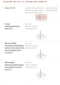
1. 0.04 s, 0.1 mV • Each of the small squares measures ____ along the x-axis (time).
• Each of the small squares measures ____ along the y-axis (ampli-
tude of the heart's electrical current).
2. P wave • No. 1 is the:
Atrial depolarization • What does it represent?
0.08-0.10 s • How long does it last?
3. PR interval (PRi) • No. 3 is the:
The onset of atrial depolar- • What does it represent?
ization to the onset of ven- • How long does it last?
tricular depolarization
0.12-0.20 s
4. QRS complex • No. 6 is the:
Ventricular depolarization • What does it represent?
(the ventricles contract) • How long does it last?
0.6-0.12 s
, nursing EKG test and i am a abnormal heart rhythm now
nursing EKG test and i am a abnormal heart rhythm now
Study online at https://quizlet.com/_hlo7dz
5. ST segment • No. 8 (which starts at the J point) is the:
The isoelectric period fol- • What does it represent?
lowing the QRS and end- • The ST segment is pathological if it depicts > ___ mV change in
ing at the beginning of the amplitude (difference of > 1 small square from the isoelectric line).
T wave, representing the
point at which both ventri-
cles are completely depo-
larized (corresponding to
the "plateau phase" of ven-
tricular action potentials).
0.1
6. 1, NSTEMI (ischemia: de- An ST segment with a depression in amplitude > 1 mm or ___ small
creased perfusion), hy- square (0.1 mV) from baseline often denotes what (2)?
pokalemia (represented by
repolarization abnormali-
ty)
7. 1, STEMI (infarction: tissue An ST segment with an elevation in amplitude > 1 mm or ___ small
death), pericarditis, coro- square (0.1 mV) from baseline often denotes what (23?
nary artery vasospasm
8. < 0.46 s Bazett's formula is a method used in electrocardiography to correct
the QT interval for heart rate, producing a corrected QT (QTc) inter-
val.
The formula is: QTc = QT / RR
Divide the QT interval time by the square root of the RR interval (the
time between two successive R-wave peaks).
, nursing EKG test and i am a abnormal heart rhythm now
nursing EKG test and i am a abnormal heart rhythm now
Study online at https://quizlet.com/_hlo7dz
A normal QTc is:
9. The time for both ventric- What does the QT interval represent?
ular depolarization and re-
polarization to occur; the
duration of an average
ventricular action poten-
tial.
10. QT interval This part of the an EKG reading represents the time for an entire
Delayed repolarization of ventricular action potential—both the depolarization and repolar-
the heart ization of the ventricles.
CVA, massive blood trans-
fusions, electrolyte deficits, What does a prolonged QTc interval ( e0.46 s) illustrate?
antiarrythmics, antibiotics
What things can cause this pattern?
11. Anticoagulants How do you treat symptomatic atrial flutter? (Hint: ABC(3)D)
Beta-blockers A:
CCBs (cardizem), Cor- B:
darone (amiodarone), Car- C (3):
dioversion D:
Digoxin
12. AV
nursing EKG test and i am a abnormal heart rhythm now
Study online at https://quizlet.com/_hlo7dz
1. 0.04 s, 0.1 mV • Each of the small squares measures ____ along the x-axis (time).
• Each of the small squares measures ____ along the y-axis (ampli-
tude of the heart's electrical current).
2. P wave • No. 1 is the:
Atrial depolarization • What does it represent?
0.08-0.10 s • How long does it last?
3. PR interval (PRi) • No. 3 is the:
The onset of atrial depolar- • What does it represent?
ization to the onset of ven- • How long does it last?
tricular depolarization
0.12-0.20 s
4. QRS complex • No. 6 is the:
Ventricular depolarization • What does it represent?
(the ventricles contract) • How long does it last?
0.6-0.12 s
, nursing EKG test and i am a abnormal heart rhythm now
nursing EKG test and i am a abnormal heart rhythm now
Study online at https://quizlet.com/_hlo7dz
5. ST segment • No. 8 (which starts at the J point) is the:
The isoelectric period fol- • What does it represent?
lowing the QRS and end- • The ST segment is pathological if it depicts > ___ mV change in
ing at the beginning of the amplitude (difference of > 1 small square from the isoelectric line).
T wave, representing the
point at which both ventri-
cles are completely depo-
larized (corresponding to
the "plateau phase" of ven-
tricular action potentials).
0.1
6. 1, NSTEMI (ischemia: de- An ST segment with a depression in amplitude > 1 mm or ___ small
creased perfusion), hy- square (0.1 mV) from baseline often denotes what (2)?
pokalemia (represented by
repolarization abnormali-
ty)
7. 1, STEMI (infarction: tissue An ST segment with an elevation in amplitude > 1 mm or ___ small
death), pericarditis, coro- square (0.1 mV) from baseline often denotes what (23?
nary artery vasospasm
8. < 0.46 s Bazett's formula is a method used in electrocardiography to correct
the QT interval for heart rate, producing a corrected QT (QTc) inter-
val.
The formula is: QTc = QT / RR
Divide the QT interval time by the square root of the RR interval (the
time between two successive R-wave peaks).
, nursing EKG test and i am a abnormal heart rhythm now
nursing EKG test and i am a abnormal heart rhythm now
Study online at https://quizlet.com/_hlo7dz
A normal QTc is:
9. The time for both ventric- What does the QT interval represent?
ular depolarization and re-
polarization to occur; the
duration of an average
ventricular action poten-
tial.
10. QT interval This part of the an EKG reading represents the time for an entire
Delayed repolarization of ventricular action potential—both the depolarization and repolar-
the heart ization of the ventricles.
CVA, massive blood trans-
fusions, electrolyte deficits, What does a prolonged QTc interval ( e0.46 s) illustrate?
antiarrythmics, antibiotics
What things can cause this pattern?
11. Anticoagulants How do you treat symptomatic atrial flutter? (Hint: ABC(3)D)
Beta-blockers A:
CCBs (cardizem), Cor- B:
darone (amiodarone), Car- C (3):
dioversion D:
Digoxin
12. AV


