

Introduction to echo
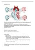
Mitral, aortic and tricuspid valves are connected to the fibrous cardiac skeleton, the pulmonary valve
is separated by a muscular sleeve.
Valvular abnormalities are typically either myxomatous (collagen degradation and elastin
fragmentation mean the valve is more prone to prolapse/ regurgitation) or fibrotic (remodelling with
calcium deposits, results in stenosis).
Aortic valve chrateristics:
3 layers:
Ventricularis – LV side made from elastic fibres
Spongiosa – in the middle, minimises friction and stress
Fibrosa – outer layer, facing the aorta, responsible for the even distribution of pressure
3 types of doppler:
1. Colour doppler
2. Pulse wave doppler – a measurement of flow at a specific point, used to measure low
velocity blood flow. This means you can quantify the speed of a signal in a particular place.
Waveforms are hollow (black) in the middle.
3. Continuous doppler – used to measure highest velocity blood flow (<6m/s) by continuously
emitting and receiving ultrasound pulses (with 2 crystals). This means that you cannot
provide an accurate location of precisely where the highest flow is occurring, only that it is
somewhere along the cursor line.
Any speed greater than 1.2m/s will case aliasing.
Mitral valve waveforms have 2 peaks – E wave and A wave. These represent passive and active
ventricular filling.
, Echo can only measure velocity, which can then be converted to pressure using the Bernoulli
equation (Pressure=4 x velocity²). There is a long and a short Bernoulli equation, you only need to
use the long Bernoulli equation is the flow before the valve is high (>1m/s). This is because you want
the difference in pressure before and after.
Aortic stenosis – affects 25% of people age 65+ years. Most common cause is degenerative in the
elderly population. Bicuspid aortic valve is a common cause of AF (1-2% of British people have a
bicuspid aortic valve). Pencil probe should be used in anyone with a suspicion of aortic stenosis,
because it has an angle of incidence of 0, so you are less likely to underestimate velocity.
Bicuspid aortic valves:
- Right-left cusp fusion (70-80%)
- Right-non cusp fusion (20-30%)
- Left-non cusp fusion (3-6%)
To classify aortic stenosis:
Bernoulli equation used to assess peak pressure gradient.
Tracing of velocity curve used to assess the mean pressure gradient.
Planimetry – performed is performed in the parasternal short axis by tracing the area of the aortic or
mitral valve while open. This represents the minimal orifice rea measure in cm².
Aortic regurgitation – aortic valve leaflets don’t meet, i.e. due to annulus dilation or leaflets
prolapsing. This results in the LV becoming volume overloaded, because blood if coming from the RA
and from the aorta. This cause myocardial fibrosis in the LV, and decreased function. To evaluate
this, you use colour doppler or colour M mode (shows the up and down motion of the heart over
time).
Mitral valve – bicuspid valve, contains an anterior and a posterior leaflet. It is supported by 2
papillary muscles and chordae tendonae.
Mitral, aortic and tricuspid valves are connected to the fibrous cardiac skeleton, the pulmonary valve
is separated by a muscular sleeve.
Valvular abnormalities are typically either myxomatous (collagen degradation and elastin
fragmentation mean the valve is more prone to prolapse/ regurgitation) or fibrotic (remodelling with
calcium deposits, results in stenosis).
Aortic valve chrateristics:
3 layers:
Ventricularis – LV side made from elastic fibres
Spongiosa – in the middle, minimises friction and stress
Fibrosa – outer layer, facing the aorta, responsible for the even distribution of pressure
3 types of doppler:
1. Colour doppler
2. Pulse wave doppler – a measurement of flow at a specific point, used to measure low
velocity blood flow. This means you can quantify the speed of a signal in a particular place.
Waveforms are hollow (black) in the middle.
3. Continuous doppler – used to measure highest velocity blood flow (<6m/s) by continuously
emitting and receiving ultrasound pulses (with 2 crystals). This means that you cannot
provide an accurate location of precisely where the highest flow is occurring, only that it is
somewhere along the cursor line.
Any speed greater than 1.2m/s will case aliasing.
Mitral valve waveforms have 2 peaks – E wave and A wave. These represent passive and active
ventricular filling.
, Echo can only measure velocity, which can then be converted to pressure using the Bernoulli
equation (Pressure=4 x velocity²). There is a long and a short Bernoulli equation, you only need to
use the long Bernoulli equation is the flow before the valve is high (>1m/s). This is because you want
the difference in pressure before and after.
Aortic stenosis – affects 25% of people age 65+ years. Most common cause is degenerative in the
elderly population. Bicuspid aortic valve is a common cause of AF (1-2% of British people have a
bicuspid aortic valve). Pencil probe should be used in anyone with a suspicion of aortic stenosis,
because it has an angle of incidence of 0, so you are less likely to underestimate velocity.
Bicuspid aortic valves:
- Right-left cusp fusion (70-80%)
- Right-non cusp fusion (20-30%)
- Left-non cusp fusion (3-6%)
To classify aortic stenosis:
Bernoulli equation used to assess peak pressure gradient.
Tracing of velocity curve used to assess the mean pressure gradient.
Planimetry – performed is performed in the parasternal short axis by tracing the area of the aortic or
mitral valve while open. This represents the minimal orifice rea measure in cm².
Aortic regurgitation – aortic valve leaflets don’t meet, i.e. due to annulus dilation or leaflets
prolapsing. This results in the LV becoming volume overloaded, because blood if coming from the RA
and from the aorta. This cause myocardial fibrosis in the LV, and decreased function. To evaluate
this, you use colour doppler or colour M mode (shows the up and down motion of the heart over
time).
Mitral valve – bicuspid valve, contains an anterior and a posterior leaflet. It is supported by 2
papillary muscles and chordae tendonae.


