
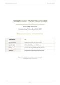
Pathophysiology Midterm Examination Jersey College 2026/2027
Pathophysiology Midterm Examination
Jersey College Study Guide
Pathophysiology Midterm Exam
150 Comprehensive Questions with Detailed Rationales
Total Questions 150
Question Format Multiple Choice (A-D), One Correct Answer
Cognitive Levels 25% Recall, 55% Application, 20% Analysis
Sections 10 Sections Covering All Pathophysiology Domains
Rationales Comprehensive with Pathophysiology Reasoning
Aligned with 2026-2027 Jersey College Curriculum Standards
and Pathophysiology Competencies
Page 1
,Pathophysiology Midterm Examination Jersey College 2026/2027
Section 1: Cellular Adaptation, Injury, and Neoplasia (Q1-25)
Q1. A 68-year-old patient with a history of prolonged immobilization following a stroke develops decreased muscle
mass in the affected limbs. Which cellular adaptation best describes this process?
A. A. Hypertrophy
B. B. Hyperplasia
C. C. Atrophy [CORRECT]
D. D. Metaplasia
Correct Answer: C
Rationale: Atrophy is a decrease in cell size that occurs in response to reduced workload, disuse, denervation, or inadequate
nutrition. In this case, prolonged immobilization leads to disuse atrophy of skeletal muscle. Hypertrophy involves an increase in
cell size, hyperplasia involves an increase in cell number, and metaplasia involves the replacement of one differentiated cell type
with another (Jersey College Pathophysiology, 2026-2027).
Q2. A 45-year-old female who is pregnant develops enlargement of the uterus. Which type of cellular adaptation is
primarily responsible for this change?
A. A. Pathologic hypertrophy
B. B. Physiologic hypertrophy combined with hyperplasia [CORRECT]
C. C. Metaplasia
D. D. Dysplasia
Correct Answer: B
Rationale: During pregnancy, the uterus undergoes both physiologic hypertrophy (increase in size of existing smooth muscle
cells) and hyperplasia (increase in number of smooth muscle cells) under the influence of estrogen and mechanical stretch. This
is a normal physiologic response, not pathologic. Metaplasia and dysplasia involve abnormal cellular changes unrelated to
normal growth adaptation (Jersey College Pathophysiology, 2026-2027).
Q3. A 52-year-old male with a long history of gastroesophageal reflux disease (GERD) is found to have columnar
epithelium replacing the normal stratified squamous epithelium in the distal esophagus. This finding is best
described as:
A. A. Dysplasia
B. B. Metaplasia [CORRECT]
C. C. Hyperplasia
D. D. Anaplasia
Correct Answer: B
Rationale: Barrett's esophagus is a classic example of metaplasia, in which the normal stratified squamous epithelium of the
esophagus is replaced by intestinal-type columnar epithelium in response to chronic acid exposure. This is a protective
adaptation, though it carries an increased risk of progression to adenocarcinoma. Dysplasia refers to abnormal cell growth with
atypical changes, hyperplasia is an increase in cell number, and anaplasia refers to undifferentiated cells seen in malignancy
(Jersey College Pathophysiology, 2026-2027).
Q4. A pathologist examines a tissue specimen and notes that cells vary in size and shape, have enlarged
hyperchromatic nuclei, and show increased mitotic figures. These changes are most consistent with:
A. A. Atrophy
Page 2
,Pathophysiology Midterm Examination Jersey College 2026/2027
B. B. Metaplasia
C. C. Dysplasia [CORRECT]
D. D. Hyperplasia
Correct Answer: C
Rationale: Dysplasia is characterized by abnormal cell growth in which cells vary in size (pleomorphism) and shape, have
enlarged hyperchromatic nuclei with increased nuclear-to-cytoplasmic ratio, and show increased mitotic figures. It represents
atypical hyperplasia that is potentially reversible if the inciting stimulus is removed, but it can also progress to cancer. Atrophy
involves cell shrinkage, metaplasia involves cell type replacement, and hyperplasia is an increase in cell number without atypical
features (Jersey College Pathophysiology, 2026-2027).
Q5. A 70-year-old male with benign prostatic hyperplasia (BPH) has an enlarged prostate gland. The cellular
adaptation responsible for this condition is:
A. A. Physiologic hyperplasia
B. B. Pathologic hyperplasia [CORRECT]
C. C. Hypertrophy only
D. D. Metaplasia
Correct Answer: B
Rationale: BPH is caused by pathologic hyperplasia, an abnormal increase in the number of cells (both glandular and stromal) in
the prostate. It is pathologic because it is not a normal physiologic response and can cause urinary obstruction. Physiologic
hyperplasia occurs in response to normal hormonal or growth factor stimulation (e.g., breast tissue during puberty). BPH
involves both hyperplasia and some degree of hypertrophy (Jersey College Pathophysiology, 2026-2027).
Q6. A patient experiences myocardial ischemia due to coronary artery occlusion. The primary mechanism by which
hypoxia causes cellular injury in this scenario is:
A. A. Activation of anaerobic metabolism leading to alkalosis
B. B. Failure of ATP-dependent sodium-potassium pump causing sodium and water influx [CORRECT]
C. C. Increased calcium efflux from the cell
D. D. Stimulation of apoptosis through intrinsic pathways
Correct Answer: B
Rationale: Hypoxia is the most common cause of cellular injury. When oxygen supply is inadequate, oxidative phosphorylation
decreases and ATP production falls. The ATP-dependent sodium-potassium pump fails, allowing sodium to accumulate
intracellularly along with water, causing cellular swelling. This is the earliest manifestation of hypoxic injury. Anaerobic
metabolism leads to lactic acidosis (not alkalosis), calcium influx (not efflux) worsens injury, and apoptosis is a later,
programmed process rather than the primary injury mechanism (Jersey College Pathophysiology, 2026-2027).
Q7. A patient with atherosclerosis experiences restoration of blood flow to a previously ischemic area of the heart,
but paradoxically, the myocardial cells show additional damage. This phenomenon is best explained by:
A. A. Free radical injury from reperfusion [CORRECT]
B. B. Coagulative necrosis from prolonged ischemia
C. C. Fat necrosis from lipase activation
D. D. Caseous necrosis from granulomatous inflammation
Correct Answer: A
Rationale: Reperfusion injury occurs when the restoration of blood flow to an ischemic area causes additional cellular damage.
The mechanism involves the generation of reactive oxygen species (ROS) by reintroduced oxygen, which overwhelms antioxidant
Page 3
, Pathophysiology Midterm Examination Jersey College 2026/2027
defenses and causes lipid peroxidation, DNA damage, and mitochondrial dysfunction. Inflammatory responses also contribute.
Coagulative necrosis occurs during the ischemic period itself, fat necrosis is specific to pancreatic injury, and caseous necrosis is
associated with tuberculosis (Jersey College Pathophysiology, 2026-2027).
Q8. Which of the following types of necrosis is most commonly associated with tuberculosis infection?
A. A. Coagulative necrosis
B. B. Liquefactive necrosis
C. C. Caseous necrosis [CORRECT]
D. D. Fat necrosis
Correct Answer: C
Rationale: Caseous necrosis is a distinctive form of necrosis most commonly associated with tuberculosis. It appears as a soft,
cheese-like (caseous) material and is a combination of coagulative and liquefactive necrosis. Microscopically, it shows
amorphous eosinophilic debris with loss of cellular detail. Coagulative necrosis is seen in heart and kidney infarcts, liquefactive
necrosis occurs in the CNS and bacterial infections, and fat necrosis occurs in the pancreas (Jersey College Pathophysiology,
2026-2027).
Q9. A 55-year-old patient presents with a brain infarct. The necrotic tissue in the brain would most likely appear as:
A. A. Firm, pale tissue with preserved cellular outlines
B. B. Soft, liquefied tissue with loss of cellular structure [CORRECT]
C. C. Cheese-like, granulomatous debris
D. D. Dry, leathery tissue with mummification
Correct Answer: B
Rationale: The central nervous system undergoes liquefactive necrosis because brain tissue is rich in lipids and has limited
connective tissue support. Unlike coagulative necrosis (which preserves cellular outlines), liquefactive necrosis results in
complete dissolution of the tissue, forming a soft, liquid-like area. This occurs because lysosomal enzymes from microglial cells
digest the necrotic tissue. Firm pale tissue describes coagulative necrosis (heart, kidney), cheese-like describes caseous necrosis
(TB), and dry leathery tissue describes dry gangrene (Jersey College Pathophysiology, 2026-2027).
Q10. A patient with acute pancreatitis develops periumbilical discoloration. This finding is most directly related to
which type of necrosis?
A. A. Coagulative necrosis
B. B. Fat necrosis [CORRECT]
C. C. Liquefactive necrosis
D. D. Gangrenous necrosis
Correct Answer: B
Rationale: Fat necrosis occurs when lipases are released from injured pancreatic cells and act on peripancreatic and abdominal
fat, splitting triglycerides into glycerol and free fatty acids. The fatty acids combine with calcium to form calcium soaps
(saponification), which appear as chalky white deposits. Cullen's sign (periumbilical discoloration) and Grey Turner's sign (flank
discoloration) are associated with hemorrhagic pancreatitis and fat necrosis. This process is distinct from coagulative,
liquefactive, and gangrenous necrosis (Jersey College Pathophysiology, 2026-2027).
Q11. A pathologist examining a tumor biopsy notes that the cells are well-differentiated, have a slow growth rate,
and are enclosed within a fibrous capsule. These findings are most characteristic of:
Page 4
Pathophysiology Midterm Examination
Jersey College Study Guide
Pathophysiology Midterm Exam
150 Comprehensive Questions with Detailed Rationales
Total Questions 150
Question Format Multiple Choice (A-D), One Correct Answer
Cognitive Levels 25% Recall, 55% Application, 20% Analysis
Sections 10 Sections Covering All Pathophysiology Domains
Rationales Comprehensive with Pathophysiology Reasoning
Aligned with 2026-2027 Jersey College Curriculum Standards
and Pathophysiology Competencies
Page 1
,Pathophysiology Midterm Examination Jersey College 2026/2027
Section 1: Cellular Adaptation, Injury, and Neoplasia (Q1-25)
Q1. A 68-year-old patient with a history of prolonged immobilization following a stroke develops decreased muscle
mass in the affected limbs. Which cellular adaptation best describes this process?
A. A. Hypertrophy
B. B. Hyperplasia
C. C. Atrophy [CORRECT]
D. D. Metaplasia
Correct Answer: C
Rationale: Atrophy is a decrease in cell size that occurs in response to reduced workload, disuse, denervation, or inadequate
nutrition. In this case, prolonged immobilization leads to disuse atrophy of skeletal muscle. Hypertrophy involves an increase in
cell size, hyperplasia involves an increase in cell number, and metaplasia involves the replacement of one differentiated cell type
with another (Jersey College Pathophysiology, 2026-2027).
Q2. A 45-year-old female who is pregnant develops enlargement of the uterus. Which type of cellular adaptation is
primarily responsible for this change?
A. A. Pathologic hypertrophy
B. B. Physiologic hypertrophy combined with hyperplasia [CORRECT]
C. C. Metaplasia
D. D. Dysplasia
Correct Answer: B
Rationale: During pregnancy, the uterus undergoes both physiologic hypertrophy (increase in size of existing smooth muscle
cells) and hyperplasia (increase in number of smooth muscle cells) under the influence of estrogen and mechanical stretch. This
is a normal physiologic response, not pathologic. Metaplasia and dysplasia involve abnormal cellular changes unrelated to
normal growth adaptation (Jersey College Pathophysiology, 2026-2027).
Q3. A 52-year-old male with a long history of gastroesophageal reflux disease (GERD) is found to have columnar
epithelium replacing the normal stratified squamous epithelium in the distal esophagus. This finding is best
described as:
A. A. Dysplasia
B. B. Metaplasia [CORRECT]
C. C. Hyperplasia
D. D. Anaplasia
Correct Answer: B
Rationale: Barrett's esophagus is a classic example of metaplasia, in which the normal stratified squamous epithelium of the
esophagus is replaced by intestinal-type columnar epithelium in response to chronic acid exposure. This is a protective
adaptation, though it carries an increased risk of progression to adenocarcinoma. Dysplasia refers to abnormal cell growth with
atypical changes, hyperplasia is an increase in cell number, and anaplasia refers to undifferentiated cells seen in malignancy
(Jersey College Pathophysiology, 2026-2027).
Q4. A pathologist examines a tissue specimen and notes that cells vary in size and shape, have enlarged
hyperchromatic nuclei, and show increased mitotic figures. These changes are most consistent with:
A. A. Atrophy
Page 2
,Pathophysiology Midterm Examination Jersey College 2026/2027
B. B. Metaplasia
C. C. Dysplasia [CORRECT]
D. D. Hyperplasia
Correct Answer: C
Rationale: Dysplasia is characterized by abnormal cell growth in which cells vary in size (pleomorphism) and shape, have
enlarged hyperchromatic nuclei with increased nuclear-to-cytoplasmic ratio, and show increased mitotic figures. It represents
atypical hyperplasia that is potentially reversible if the inciting stimulus is removed, but it can also progress to cancer. Atrophy
involves cell shrinkage, metaplasia involves cell type replacement, and hyperplasia is an increase in cell number without atypical
features (Jersey College Pathophysiology, 2026-2027).
Q5. A 70-year-old male with benign prostatic hyperplasia (BPH) has an enlarged prostate gland. The cellular
adaptation responsible for this condition is:
A. A. Physiologic hyperplasia
B. B. Pathologic hyperplasia [CORRECT]
C. C. Hypertrophy only
D. D. Metaplasia
Correct Answer: B
Rationale: BPH is caused by pathologic hyperplasia, an abnormal increase in the number of cells (both glandular and stromal) in
the prostate. It is pathologic because it is not a normal physiologic response and can cause urinary obstruction. Physiologic
hyperplasia occurs in response to normal hormonal or growth factor stimulation (e.g., breast tissue during puberty). BPH
involves both hyperplasia and some degree of hypertrophy (Jersey College Pathophysiology, 2026-2027).
Q6. A patient experiences myocardial ischemia due to coronary artery occlusion. The primary mechanism by which
hypoxia causes cellular injury in this scenario is:
A. A. Activation of anaerobic metabolism leading to alkalosis
B. B. Failure of ATP-dependent sodium-potassium pump causing sodium and water influx [CORRECT]
C. C. Increased calcium efflux from the cell
D. D. Stimulation of apoptosis through intrinsic pathways
Correct Answer: B
Rationale: Hypoxia is the most common cause of cellular injury. When oxygen supply is inadequate, oxidative phosphorylation
decreases and ATP production falls. The ATP-dependent sodium-potassium pump fails, allowing sodium to accumulate
intracellularly along with water, causing cellular swelling. This is the earliest manifestation of hypoxic injury. Anaerobic
metabolism leads to lactic acidosis (not alkalosis), calcium influx (not efflux) worsens injury, and apoptosis is a later,
programmed process rather than the primary injury mechanism (Jersey College Pathophysiology, 2026-2027).
Q7. A patient with atherosclerosis experiences restoration of blood flow to a previously ischemic area of the heart,
but paradoxically, the myocardial cells show additional damage. This phenomenon is best explained by:
A. A. Free radical injury from reperfusion [CORRECT]
B. B. Coagulative necrosis from prolonged ischemia
C. C. Fat necrosis from lipase activation
D. D. Caseous necrosis from granulomatous inflammation
Correct Answer: A
Rationale: Reperfusion injury occurs when the restoration of blood flow to an ischemic area causes additional cellular damage.
The mechanism involves the generation of reactive oxygen species (ROS) by reintroduced oxygen, which overwhelms antioxidant
Page 3
, Pathophysiology Midterm Examination Jersey College 2026/2027
defenses and causes lipid peroxidation, DNA damage, and mitochondrial dysfunction. Inflammatory responses also contribute.
Coagulative necrosis occurs during the ischemic period itself, fat necrosis is specific to pancreatic injury, and caseous necrosis is
associated with tuberculosis (Jersey College Pathophysiology, 2026-2027).
Q8. Which of the following types of necrosis is most commonly associated with tuberculosis infection?
A. A. Coagulative necrosis
B. B. Liquefactive necrosis
C. C. Caseous necrosis [CORRECT]
D. D. Fat necrosis
Correct Answer: C
Rationale: Caseous necrosis is a distinctive form of necrosis most commonly associated with tuberculosis. It appears as a soft,
cheese-like (caseous) material and is a combination of coagulative and liquefactive necrosis. Microscopically, it shows
amorphous eosinophilic debris with loss of cellular detail. Coagulative necrosis is seen in heart and kidney infarcts, liquefactive
necrosis occurs in the CNS and bacterial infections, and fat necrosis occurs in the pancreas (Jersey College Pathophysiology,
2026-2027).
Q9. A 55-year-old patient presents with a brain infarct. The necrotic tissue in the brain would most likely appear as:
A. A. Firm, pale tissue with preserved cellular outlines
B. B. Soft, liquefied tissue with loss of cellular structure [CORRECT]
C. C. Cheese-like, granulomatous debris
D. D. Dry, leathery tissue with mummification
Correct Answer: B
Rationale: The central nervous system undergoes liquefactive necrosis because brain tissue is rich in lipids and has limited
connective tissue support. Unlike coagulative necrosis (which preserves cellular outlines), liquefactive necrosis results in
complete dissolution of the tissue, forming a soft, liquid-like area. This occurs because lysosomal enzymes from microglial cells
digest the necrotic tissue. Firm pale tissue describes coagulative necrosis (heart, kidney), cheese-like describes caseous necrosis
(TB), and dry leathery tissue describes dry gangrene (Jersey College Pathophysiology, 2026-2027).
Q10. A patient with acute pancreatitis develops periumbilical discoloration. This finding is most directly related to
which type of necrosis?
A. A. Coagulative necrosis
B. B. Fat necrosis [CORRECT]
C. C. Liquefactive necrosis
D. D. Gangrenous necrosis
Correct Answer: B
Rationale: Fat necrosis occurs when lipases are released from injured pancreatic cells and act on peripancreatic and abdominal
fat, splitting triglycerides into glycerol and free fatty acids. The fatty acids combine with calcium to form calcium soaps
(saponification), which appear as chalky white deposits. Cullen's sign (periumbilical discoloration) and Grey Turner's sign (flank
discoloration) are associated with hemorrhagic pancreatitis and fat necrosis. This process is distinct from coagulative,
liquefactive, and gangrenous necrosis (Jersey College Pathophysiology, 2026-2027).
Q11. A pathologist examining a tumor biopsy notes that the cells are well-differentiated, have a slow growth rate,
and are enclosed within a fibrous capsule. These findings are most characteristic of:
Page 4


