
BIOL251 Human Anatomy & Physiology I w/Lab (2026) |
Module 1 Exam Review | 100 Questions & Answers Summary
| Ultimate Exam Mastery Guide | High-Yield Revision
Resource | Portage Learning
,Question 1
According to the image, which of the following correctly lists the four processes of respiration in the
correct sequence?
□ Cellular respiration → Internal respiration → External respiration → Breathing
☑ Breathing → External respiration → Internal respiration → Cellular respiration
□ Breathing → Internal respiration → External respiration → Cellular respiration
□ External respiration → Breathing → Cellular respiration → Internal respiration
Answer: Breathing → External respiration → Internal respiration → Cellular respiration
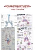
,Rationale: The image clearly outlines the four-step process in this specific order: Breathing (exchange
between atmosphere and lungs) → External respiration (exchange between lungs and blood) → Internal
respiration (exchange between blood and body cells) → Cellular respiration (use of oxygen by cells to
release energy). This is the logical cascade from the environment to the cellular level.
Question 2
Which of the following best describes the process of external respiration as defined in the image?
□ Intake of oxygen and release of carbon dioxide at the cellular level
□ Movement of air into and out of the lungs
☑ Exchange of gases between the lungs and the blood
□ Use of oxygen by cells to release energy
Answer: Exchange of gases between the lungs and the blood
Rationale: The image explicitly defines external respiration as the "Exchange of gases between lungs and
blood." This occurs at the alveoli-capillary membrane where oxygen diffuses into the blood and carbon
dioxide diffuses out of the blood to be exhaled.
Question 3
Which of the following structures is NOT listed as a main part of the respiratory system in the image?
□ Trachea
□ Alveoli
☑ Esophagus
□ Diaphragm
Answer: Esophagus
Rationale: The image lists the main parts as: Nasal cavity, Pharynx, Larynx, Trachea, Bronchi,
Bronchioles, Alveoli, Lungs, and Diaphragm. The esophagus is part of the digestive system (though it
shares the pharyngeal region with the respiratory tract), and it is notably absent from the listed
structures.
, Question 4
During inhalation, which of the following correctly describes the movement of the diaphragm and the
resulting change in the chest cavity?
☑ Diaphragm contracts (moves down); chest cavity expands
□ Diaphragm relaxes (moves up); chest cavity shrinks
□ Diaphragm contracts (moves down); chest cavity shrinks
□ Diaphragm relaxes (moves up); chest cavity expands
Answer: Diaphragm contracts (moves down); chest cavity expands
Rationale: The image's "Breathing mechanism" section clearly states: Inhalation → Diaphragm contracts
(moves down) → Chest cavity expands → Air enters lungs. The contraction of the diaphragm flattens it,
increasing the thoracic volume and decreasing intrathoracic pressure, drawing air into the lungs.
Question 5
The process of cellular respiration, as defined in the image, serves what primary purpose in the body?
□ Exchange of gases between blood and body cells
□ Movement of air through the conducting airways
☑ Use of oxygen by cells to release energy
□ Removal of carbon dioxide from the alveoli
Answer: Use of oxygen by cells to release energy
Rationale: The image explicitly describes cellular respiration as the "Use of oxygen by cells to release
energy." This is the metabolic process (aerobic respiration) occurring within mitochondria that produces
ATP from glucose and oxygen, with carbon dioxide as a waste product.
Question 6
Which of the following correctly traces the pathway of air from the external environment to the alveoli,
according to the structures listed in the image?
Module 1 Exam Review | 100 Questions & Answers Summary
| Ultimate Exam Mastery Guide | High-Yield Revision
Resource | Portage Learning
,Question 1
According to the image, which of the following correctly lists the four processes of respiration in the
correct sequence?
□ Cellular respiration → Internal respiration → External respiration → Breathing
☑ Breathing → External respiration → Internal respiration → Cellular respiration
□ Breathing → Internal respiration → External respiration → Cellular respiration
□ External respiration → Breathing → Cellular respiration → Internal respiration
Answer: Breathing → External respiration → Internal respiration → Cellular respiration
,Rationale: The image clearly outlines the four-step process in this specific order: Breathing (exchange
between atmosphere and lungs) → External respiration (exchange between lungs and blood) → Internal
respiration (exchange between blood and body cells) → Cellular respiration (use of oxygen by cells to
release energy). This is the logical cascade from the environment to the cellular level.
Question 2
Which of the following best describes the process of external respiration as defined in the image?
□ Intake of oxygen and release of carbon dioxide at the cellular level
□ Movement of air into and out of the lungs
☑ Exchange of gases between the lungs and the blood
□ Use of oxygen by cells to release energy
Answer: Exchange of gases between the lungs and the blood
Rationale: The image explicitly defines external respiration as the "Exchange of gases between lungs and
blood." This occurs at the alveoli-capillary membrane where oxygen diffuses into the blood and carbon
dioxide diffuses out of the blood to be exhaled.
Question 3
Which of the following structures is NOT listed as a main part of the respiratory system in the image?
□ Trachea
□ Alveoli
☑ Esophagus
□ Diaphragm
Answer: Esophagus
Rationale: The image lists the main parts as: Nasal cavity, Pharynx, Larynx, Trachea, Bronchi,
Bronchioles, Alveoli, Lungs, and Diaphragm. The esophagus is part of the digestive system (though it
shares the pharyngeal region with the respiratory tract), and it is notably absent from the listed
structures.
, Question 4
During inhalation, which of the following correctly describes the movement of the diaphragm and the
resulting change in the chest cavity?
☑ Diaphragm contracts (moves down); chest cavity expands
□ Diaphragm relaxes (moves up); chest cavity shrinks
□ Diaphragm contracts (moves down); chest cavity shrinks
□ Diaphragm relaxes (moves up); chest cavity expands
Answer: Diaphragm contracts (moves down); chest cavity expands
Rationale: The image's "Breathing mechanism" section clearly states: Inhalation → Diaphragm contracts
(moves down) → Chest cavity expands → Air enters lungs. The contraction of the diaphragm flattens it,
increasing the thoracic volume and decreasing intrathoracic pressure, drawing air into the lungs.
Question 5
The process of cellular respiration, as defined in the image, serves what primary purpose in the body?
□ Exchange of gases between blood and body cells
□ Movement of air through the conducting airways
☑ Use of oxygen by cells to release energy
□ Removal of carbon dioxide from the alveoli
Answer: Use of oxygen by cells to release energy
Rationale: The image explicitly describes cellular respiration as the "Use of oxygen by cells to release
energy." This is the metabolic process (aerobic respiration) occurring within mitochondria that produces
ATP from glucose and oxygen, with carbon dioxide as a waste product.
Question 6
Which of the following correctly traces the pathway of air from the external environment to the alveoli,
according to the structures listed in the image?


