Anatomy & Physiology — Skeletal System (With
Pictures)
Complete Exam Pack · 110 Questions
BIOD 151 — Essential Human Anatomy & Physiology I with Lab | Skeletal System Comprehensive Examination
| Updated 2026/2027
Bone Tissue Histology & Development · Axial & Appendicular Skeleton · Bone Markings & Landmarks · Joint
Classifications & Movements
Radiographic Identification · Clinical Correlations & Skeletal Pathologies · Laboratory Applications in
Osteology
Undergraduate Health Sciences | Respondus LockDown Browser Secure Exam Format | 110 Questions · 100
Points · 90 Minutes
Exam Structure
The Anatomy & Physiology Skeletal System Examination for the 2026/2027 academic cycle is a
110-question secure online examination administered through the Respondus LockDown
Browser with webcam monitoring. The instrument blends multiple-choice, multiple-select, short-
answer, image-labeling, and figure-identification items across a 90-minute window for 100 total
points. Item weighting below reflects the distribution used throughout this pack.
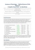
Section Items Weighting Points
I — Bone Tissue, Histology & Development Q1–Q20 20 items × 0.5 pt 10 pts
II — Axial Skeleton Q21–Q38 18 items × 0.5 pt 9 pts
III — Appendicular Skeleton Q39–Q54 16 items × 0.5 pt 8 pts
IV — Joints & Movements Q55–Q70 16 items × 0.5 pt 8 pts
V — Bone Markings, Radiology & Pathology Q71–Q84 14 items × 0.5 pt 7 pts
VI — Multiple Select Q85–Q90 6 items × 1 pt 6 pts
VII — Figure Identification (9 labeled plates) Q91–Q99 9 items, 6–14 pts 94 pts*
VIII — Short Answer Q100–Q106 7 items × 3 pts 21 pts*
IX — Clinical Correlation Q107–Q110 4 items, 2–3 pts 10 pts*
TOTAL (scaled) 110 items — 100 pts
*Raw points across all nine sections total 173 and are scaled to the reported 100-point maximum
(multiply each raw score by 0.578). The 110 items comprise 84 multiple-choice items, 6 multiple-
select items, 9 figure-identification plates carrying 129 individually labeled structures, 7 short-
answer items, and 4 clinical correlations.
Introduction
This guide prepares undergraduate health sciences students on the structural organization,
anatomical landmarks, and physiological functions of the human skeletal system. Content
emphasizes visual identification of bones and markings, classification of joints and movements,
histological understanding of bone tissue, and clinical correlations aligned with Portage Learning
BIOD 151 course objectives, ensuring learners can accurately label skeletal structures on
Portage Learning · BIOD 151 Essential Human Anatomy & Physiology I with Lab · Skeletal System Exam Pack · 2026–2027
,diagrams, explain biomechanical relationships, interpret radiographic images, and apply
foundational knowledge to musculoskeletal case scenarios.
Section I · Bone Tissue, Histology & Development (Q1–Q20)
Question 1 (0.5 points · Multiple Choice)
Which of the following is NOT a primary function of the skeletal system?
A. Support and protection of soft organs
B. Storage of calcium and phosphate
C. Production of blood cells (hematopoiesis)
D. Secretion of digestive enzymes ✓
Correct Answer: D. Secretion of digestive enzymes
Rationale: The skeleton supports the body, protects organs such as the brain and heart, stores minerals
(chiefly calcium and phosphate) and triglycerides, houses hematopoietic red marrow, and provides levers for
muscle-driven movement. Digestive enzyme secretion is a function of the pancreas, stomach, and salivary
glands and has no skeletal component.
References: Portage Learning BIOD 151 skeletal module and Lab 4 content; Marieb & Hoehn, Human Anatomy &
Physiology, 11th ed., Ch. 6; Saladin, Anatomy & Physiology, 9th ed. Ch. 7.
Question 2 (0.5 points · Multiple Choice)
How many bones are in the typical adult human skeleton?
A. 180
B. 196
C. 206 ✓
D. 270
Correct Answer: C. 206
Rationale: The adult skeleton contains 206 named bones: 80 axial and 126 appendicular. Infants are born
with roughly 270 bones, many of which fuse during growth — for example the sacral and coccygeal vertebrae
and the three components of each coxal bone — which is why option D describes the newborn rather than the
adult count.
References: Portage Learning BIOD 151 skeletal module and Lab 4 content; Marieb & Hoehn, 11th ed., Ch. 7; Martini,
Fundamentals of Anatomy & Physiology, 11th ed. Ch. 7.
Question 3 (0.5 points · Multiple Choice)
Which bone is classified as a long bone?
A. Carpal
B. Phalanx ✓
C. Patella
D. Vertebra
Correct Answer: B. Phalanx
Rationale: Long bones are longer than they are wide and possess a shaft with two expanded ends; the
phalanges qualify despite their small size, as do the femur, humerus, radius, ulna, tibia, fibula, metacarpals,
Portage Learning · BIOD 151 Essential Human Anatomy & Physiology I with Lab · Skeletal System Exam Pack · 2026–2027
,and metatarsals. Carpals are short bones, the patella is a sesamoid bone, and vertebrae are irregular bones.
Classification depends on shape and proportion, not absolute length.
References: Portage Learning BIOD 151 skeletal module and Lab 4 content; Marieb & Hoehn, Human Anatomy &
Physiology, 11th ed., Ch. 6; Saladin, Anatomy & Physiology, 9th ed. Ch. 7.
Question 4 (0.5 points · Multiple Choice)
The structural and functional unit of compact bone is the:
A. Trabecula
B. Osteon (Haversian system) ✓
C. Lacuna
D. Epiphysis
Correct Answer: B. Osteon (Haversian system)
Rationale: Compact bone is built from osteons — cylindrical units of concentric lamellae surrounding a
central (Haversian) canal that carries blood vessels and nerves. Trabeculae are the strut-like elements of
spongy bone, lacunae are the tiny cavities housing individual osteocytes, and the epiphysis is a gross region of a
long bone rather than a microscopic unit.
References: Portage Learning BIOD 151 skeletal module and Lab 4 content; Marieb & Hoehn, Human Anatomy &
Physiology, 11th ed., Ch. 6; Martini, Fundamentals of Anatomy & Physiology, 11th ed. Ch. 6.
Question 5 (0.5 points · Multiple Choice)
Mature bone cells that maintain bone matrix are called ________ and they occupy small cavities
called ________.
A. osteoblasts; canaliculi
B. osteoclasts; lacunae
C. osteocytes; lacunae ✓
D. osteocytes; trabeculae
Correct Answer: C. osteocytes; lacunae
Rationale: Osteocytes are mature bone cells trapped within lacunae in the calcified matrix; they monitor
and maintain the matrix and signal for repair. Osteoblasts are the bone-building cells that become osteocytes
once surrounded by matrix, osteoclasts are the large multinucleated cells that resorb bone, and canaliculi are
the tiny channels connecting lacunae rather than the cavities themselves.
References: Portage Learning BIOD 151 skeletal module and Lab 4 content; Marieb & Hoehn, Human Anatomy &
Physiology, 11th ed., Ch. 6; Saladin, Anatomy & Physiology, 9th ed. Ch. 7.
Question 6 (0.5 points · Multiple Choice)
What is the function of canaliculi in compact bone?
A. They store yellow marrow.
B. They connect lacunae, allowing nutrients and wastes to pass between osteocytes
and the central canal. ✓
C. They carry lymph away from the bone surface.
D. They anchor tendons to the periosteum.
Correct Answer: B. They connect lacunae, allowing nutrients and wastes to pass between
osteocytes and the central canal.
Portage Learning · BIOD 151 Essential Human Anatomy & Physiology I with Lab · Skeletal System Exam Pack · 2026–2027
, Rationale: Canaliculi are hairlike channels radiating from each lacuna. Osteocyte processes extend through
them and form gap junctions with neighbors, creating a continuous route for nutrients and waste between the
central canal blood supply and cells buried in calcified matrix — the reason osteocytes survive despite being
fully entombed.
References: Portage Learning BIOD 151 skeletal module and Lab 4 content; Marieb & Hoehn, Human Anatomy &
Physiology, 11th ed., Ch. 6; Martini, Fundamentals of Anatomy & Physiology, 11th ed. Ch. 6.
Question 7 (0.5 points · Multiple Choice)
The central (Haversian) canal of an osteon contains:
A. Yellow marrow only
B. Blood vessels and nerves ✓
C. Hyaline cartilage
D. Osteoclasts exclusively
Correct Answer: B. Blood vessels and nerves
Rationale: Each osteon is organized around a central canal carrying small blood vessels and nerve fibers
that supply the surrounding lamellae. Yellow marrow occupies the medullary cavity of the diaphysis, hyaline
cartilage covers articular surfaces, and osteoclasts are found chiefly at resorption surfaces such as the
endosteum.
References: Portage Learning BIOD 151 skeletal module and Lab 4 content; Marieb & Hoehn, Human Anatomy &
Physiology, 11th ed., Ch. 6; Saladin, Anatomy & Physiology, 9th ed. Ch. 7.
Question 8 (0.5 points · Multiple Choice)
Perforating (Volkmann's) canals differ from central canals in that they:
A. Run parallel to the long axis of the bone
B. Run perpendicular to the long axis, connecting the periosteum with central canals
✓
C. Contain only lymphatic vessels
D. Are found only in spongy bone
Correct Answer: B. Run perpendicular to the long axis, connecting the periosteum with
central canals
Rationale: Perforating canals travel at right angles to the shaft, linking the blood and nerve supply of the
periosteum with the central canals and the medullary cavity. Central canals are the ones oriented parallel to
the long axis. Both canal systems belong to compact bone; spongy bone has no osteons of this kind because its
trabeculae are nourished directly by marrow.
References: Portage Learning BIOD 151 skeletal module and Lab 4 content; Marieb & Hoehn, Human Anatomy &
Physiology, 11th ed., Ch. 6; Martini, Fundamentals of Anatomy & Physiology, 11th ed. Ch. 6.
Question 9 (0.5 points · Multiple Choice)
Osteoblasts are best described as:
A. Bone-resorbing cells derived from monocytes
B. Bone-forming cells that secrete osteoid ✓
C. Mature cells within lacunae
D. Stem cells of the endosteum
Correct Answer: B. Bone-forming cells that secrete osteoid
Portage Learning · BIOD 151 Essential Human Anatomy & Physiology I with Lab · Skeletal System Exam Pack · 2026–2027
Pictures)
Complete Exam Pack · 110 Questions
BIOD 151 — Essential Human Anatomy & Physiology I with Lab | Skeletal System Comprehensive Examination
| Updated 2026/2027
Bone Tissue Histology & Development · Axial & Appendicular Skeleton · Bone Markings & Landmarks · Joint
Classifications & Movements
Radiographic Identification · Clinical Correlations & Skeletal Pathologies · Laboratory Applications in
Osteology
Undergraduate Health Sciences | Respondus LockDown Browser Secure Exam Format | 110 Questions · 100
Points · 90 Minutes
Exam Structure
The Anatomy & Physiology Skeletal System Examination for the 2026/2027 academic cycle is a
110-question secure online examination administered through the Respondus LockDown
Browser with webcam monitoring. The instrument blends multiple-choice, multiple-select, short-
answer, image-labeling, and figure-identification items across a 90-minute window for 100 total
points. Item weighting below reflects the distribution used throughout this pack.
Section Items Weighting Points
I — Bone Tissue, Histology & Development Q1–Q20 20 items × 0.5 pt 10 pts
II — Axial Skeleton Q21–Q38 18 items × 0.5 pt 9 pts
III — Appendicular Skeleton Q39–Q54 16 items × 0.5 pt 8 pts
IV — Joints & Movements Q55–Q70 16 items × 0.5 pt 8 pts
V — Bone Markings, Radiology & Pathology Q71–Q84 14 items × 0.5 pt 7 pts
VI — Multiple Select Q85–Q90 6 items × 1 pt 6 pts
VII — Figure Identification (9 labeled plates) Q91–Q99 9 items, 6–14 pts 94 pts*
VIII — Short Answer Q100–Q106 7 items × 3 pts 21 pts*
IX — Clinical Correlation Q107–Q110 4 items, 2–3 pts 10 pts*
TOTAL (scaled) 110 items — 100 pts
*Raw points across all nine sections total 173 and are scaled to the reported 100-point maximum
(multiply each raw score by 0.578). The 110 items comprise 84 multiple-choice items, 6 multiple-
select items, 9 figure-identification plates carrying 129 individually labeled structures, 7 short-
answer items, and 4 clinical correlations.
Introduction
This guide prepares undergraduate health sciences students on the structural organization,
anatomical landmarks, and physiological functions of the human skeletal system. Content
emphasizes visual identification of bones and markings, classification of joints and movements,
histological understanding of bone tissue, and clinical correlations aligned with Portage Learning
BIOD 151 course objectives, ensuring learners can accurately label skeletal structures on
Portage Learning · BIOD 151 Essential Human Anatomy & Physiology I with Lab · Skeletal System Exam Pack · 2026–2027
,diagrams, explain biomechanical relationships, interpret radiographic images, and apply
foundational knowledge to musculoskeletal case scenarios.
Section I · Bone Tissue, Histology & Development (Q1–Q20)
Question 1 (0.5 points · Multiple Choice)
Which of the following is NOT a primary function of the skeletal system?
A. Support and protection of soft organs
B. Storage of calcium and phosphate
C. Production of blood cells (hematopoiesis)
D. Secretion of digestive enzymes ✓
Correct Answer: D. Secretion of digestive enzymes
Rationale: The skeleton supports the body, protects organs such as the brain and heart, stores minerals
(chiefly calcium and phosphate) and triglycerides, houses hematopoietic red marrow, and provides levers for
muscle-driven movement. Digestive enzyme secretion is a function of the pancreas, stomach, and salivary
glands and has no skeletal component.
References: Portage Learning BIOD 151 skeletal module and Lab 4 content; Marieb & Hoehn, Human Anatomy &
Physiology, 11th ed., Ch. 6; Saladin, Anatomy & Physiology, 9th ed. Ch. 7.
Question 2 (0.5 points · Multiple Choice)
How many bones are in the typical adult human skeleton?
A. 180
B. 196
C. 206 ✓
D. 270
Correct Answer: C. 206
Rationale: The adult skeleton contains 206 named bones: 80 axial and 126 appendicular. Infants are born
with roughly 270 bones, many of which fuse during growth — for example the sacral and coccygeal vertebrae
and the three components of each coxal bone — which is why option D describes the newborn rather than the
adult count.
References: Portage Learning BIOD 151 skeletal module and Lab 4 content; Marieb & Hoehn, 11th ed., Ch. 7; Martini,
Fundamentals of Anatomy & Physiology, 11th ed. Ch. 7.
Question 3 (0.5 points · Multiple Choice)
Which bone is classified as a long bone?
A. Carpal
B. Phalanx ✓
C. Patella
D. Vertebra
Correct Answer: B. Phalanx
Rationale: Long bones are longer than they are wide and possess a shaft with two expanded ends; the
phalanges qualify despite their small size, as do the femur, humerus, radius, ulna, tibia, fibula, metacarpals,
Portage Learning · BIOD 151 Essential Human Anatomy & Physiology I with Lab · Skeletal System Exam Pack · 2026–2027
,and metatarsals. Carpals are short bones, the patella is a sesamoid bone, and vertebrae are irregular bones.
Classification depends on shape and proportion, not absolute length.
References: Portage Learning BIOD 151 skeletal module and Lab 4 content; Marieb & Hoehn, Human Anatomy &
Physiology, 11th ed., Ch. 6; Saladin, Anatomy & Physiology, 9th ed. Ch. 7.
Question 4 (0.5 points · Multiple Choice)
The structural and functional unit of compact bone is the:
A. Trabecula
B. Osteon (Haversian system) ✓
C. Lacuna
D. Epiphysis
Correct Answer: B. Osteon (Haversian system)
Rationale: Compact bone is built from osteons — cylindrical units of concentric lamellae surrounding a
central (Haversian) canal that carries blood vessels and nerves. Trabeculae are the strut-like elements of
spongy bone, lacunae are the tiny cavities housing individual osteocytes, and the epiphysis is a gross region of a
long bone rather than a microscopic unit.
References: Portage Learning BIOD 151 skeletal module and Lab 4 content; Marieb & Hoehn, Human Anatomy &
Physiology, 11th ed., Ch. 6; Martini, Fundamentals of Anatomy & Physiology, 11th ed. Ch. 6.
Question 5 (0.5 points · Multiple Choice)
Mature bone cells that maintain bone matrix are called ________ and they occupy small cavities
called ________.
A. osteoblasts; canaliculi
B. osteoclasts; lacunae
C. osteocytes; lacunae ✓
D. osteocytes; trabeculae
Correct Answer: C. osteocytes; lacunae
Rationale: Osteocytes are mature bone cells trapped within lacunae in the calcified matrix; they monitor
and maintain the matrix and signal for repair. Osteoblasts are the bone-building cells that become osteocytes
once surrounded by matrix, osteoclasts are the large multinucleated cells that resorb bone, and canaliculi are
the tiny channels connecting lacunae rather than the cavities themselves.
References: Portage Learning BIOD 151 skeletal module and Lab 4 content; Marieb & Hoehn, Human Anatomy &
Physiology, 11th ed., Ch. 6; Saladin, Anatomy & Physiology, 9th ed. Ch. 7.
Question 6 (0.5 points · Multiple Choice)
What is the function of canaliculi in compact bone?
A. They store yellow marrow.
B. They connect lacunae, allowing nutrients and wastes to pass between osteocytes
and the central canal. ✓
C. They carry lymph away from the bone surface.
D. They anchor tendons to the periosteum.
Correct Answer: B. They connect lacunae, allowing nutrients and wastes to pass between
osteocytes and the central canal.
Portage Learning · BIOD 151 Essential Human Anatomy & Physiology I with Lab · Skeletal System Exam Pack · 2026–2027
, Rationale: Canaliculi are hairlike channels radiating from each lacuna. Osteocyte processes extend through
them and form gap junctions with neighbors, creating a continuous route for nutrients and waste between the
central canal blood supply and cells buried in calcified matrix — the reason osteocytes survive despite being
fully entombed.
References: Portage Learning BIOD 151 skeletal module and Lab 4 content; Marieb & Hoehn, Human Anatomy &
Physiology, 11th ed., Ch. 6; Martini, Fundamentals of Anatomy & Physiology, 11th ed. Ch. 6.
Question 7 (0.5 points · Multiple Choice)
The central (Haversian) canal of an osteon contains:
A. Yellow marrow only
B. Blood vessels and nerves ✓
C. Hyaline cartilage
D. Osteoclasts exclusively
Correct Answer: B. Blood vessels and nerves
Rationale: Each osteon is organized around a central canal carrying small blood vessels and nerve fibers
that supply the surrounding lamellae. Yellow marrow occupies the medullary cavity of the diaphysis, hyaline
cartilage covers articular surfaces, and osteoclasts are found chiefly at resorption surfaces such as the
endosteum.
References: Portage Learning BIOD 151 skeletal module and Lab 4 content; Marieb & Hoehn, Human Anatomy &
Physiology, 11th ed., Ch. 6; Saladin, Anatomy & Physiology, 9th ed. Ch. 7.
Question 8 (0.5 points · Multiple Choice)
Perforating (Volkmann's) canals differ from central canals in that they:
A. Run parallel to the long axis of the bone
B. Run perpendicular to the long axis, connecting the periosteum with central canals
✓
C. Contain only lymphatic vessels
D. Are found only in spongy bone
Correct Answer: B. Run perpendicular to the long axis, connecting the periosteum with
central canals
Rationale: Perforating canals travel at right angles to the shaft, linking the blood and nerve supply of the
periosteum with the central canals and the medullary cavity. Central canals are the ones oriented parallel to
the long axis. Both canal systems belong to compact bone; spongy bone has no osteons of this kind because its
trabeculae are nourished directly by marrow.
References: Portage Learning BIOD 151 skeletal module and Lab 4 content; Marieb & Hoehn, Human Anatomy &
Physiology, 11th ed., Ch. 6; Martini, Fundamentals of Anatomy & Physiology, 11th ed. Ch. 6.
Question 9 (0.5 points · Multiple Choice)
Osteoblasts are best described as:
A. Bone-resorbing cells derived from monocytes
B. Bone-forming cells that secrete osteoid ✓
C. Mature cells within lacunae
D. Stem cells of the endosteum
Correct Answer: B. Bone-forming cells that secrete osteoid
Portage Learning · BIOD 151 Essential Human Anatomy & Physiology I with Lab · Skeletal System Exam Pack · 2026–2027