
NURSING
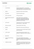
# Term Definition
1 3 muscle layers Endocarium Myocardium – muscle Ex. MI =
myocardial infarction, to the muscle Epicardium
Heart pumps blood to the body with the muscle
2 Septum Separates the right and left chambers
3 Pericardium Visceral and parietal layers
4 Fluid in pericardium Heart gets squished
5 How blood flows through the Heart sup/inf vena cava right atrium
heart tricuspid valve right ventricle pulmonary
artery (carries deoxygenated blood) lungs
(oxygenated) pulmonary vein left atrium
left ventricle aorta the body
6 Left ventricle Pumps blood to the rest of the body Problem
with left ventricle = cardiac output
7 Mechanical system Diastole Systole Cardiac output
8 Diastole Myocardium relaxes Ventricles fill back up
9 Systole Contraction of the myocardium Blood is ejected
from the ventricles
10 Cardiac output Amount of blood the heart pumps in 1 min
Usually 4-8 L, depending on patient average is 5
L
11 Myocardium Coronary circulation
12 2 major arteries fill during Left: provides 65-85% of blood supply to
diastole myocardium. Branches into left ascending – LAD
and left circumflex Right: right coronary artery –
RCA
13 Collateral circulation Development of arterial branching that occurs
within the coronary circulation - when occlusion
of the coronary arteries occurs slowly over a
long period
,NURSING
# Term Definition
14 Valves – 4 that keep blood Atrioventricular Semilunar Pulmonic Aortic
flowing in forward direction
15 Atrioventricular Valve Mitral (Bicuspid): between left atrium and left
ventricle Tricuspid: between the right atrium
and right ventricle
16 Pulmonic Valve Between right ventricle artery
17 Aortic Valve Between left ventricle artery
18 Conduction system Specialized nerves tissue SA node AV node
right and left bundles ventricle contraction
Electrical conduction
19 Specialized nerves tissue depolarization and cardiac circulation
20 Electrical conduction Detected on the body surface through electrode
recorded on telemetry monitor and EKG/ECG
21 Physiologic Changes with Aging Left ventricular hypertrophy (LVH) in systolic
-Heart blood pressure, vascular changes
22 Physiologic Changes with Aging Degenerative changes in vessel walls affecting
-Arteries blood flow- atherosclerosis
23 Physiologic Changes with Aging - Prolonged atrioventricular (AV) conduction, rate,
Conduction system and rhythm disturbances
24 Physiologic Changes with Aging - Decreased rate response with increased cardiac
Exercise and cardiovascular workload Exercise max heart rate Ex. 80 yr old
response on treadmill, heart rate will not get as high as
young person
25 Cardiac Assessment Past medical history Medications current and
past Aspirin: prevents platelets from clumping
Surgery- especially for cardiac issues Family
history
,NURSING
# Term Definition
26 Risk Factors -Modifiable Hypertension Diabetes Cholesterol levels
Obesity (metabolic syndrome) Diet Smoking:
restricts blood vessels Sedentary lifestyle
ETOH/drug abuse Socioeconomic status Stress
( SNS): related to catecholamines, HR, BP
Depression Periodontal disease: bad teeth leads
to endocarditis
27 Risk Factors - Non-Modifiable Age Gender Ethnicity Genetics (family history)
28 Indicators of cardiac function and Level of consciousness: awake, alert, agitated,
cardiac output confused What is their orientation? Skin
temperature: warm, dry, diaphoretic
Diaphoretic = pale, clammy Capillary refill: 2
seconds or delayed Pulse rate and volume:
strong, weak, thready Thready = hard to palpate
Urine output: at least 30 mL/hr
29 Cardiac Signs and Symptoms - Most common complaint in acute myocardial
Chest Pain infarction (AMI) Location, duration, radiation to
arm, jaw, back-scapula Other signs and
symptoms: nausea, diaphoresis, fatigue Use
pain scale to rate 0 – 10 Very important to ask if
pain is getting better Want pain to be 0 =
indicates NO ischemia Always assess pain, don’t
assume etiology Chest pain that occurs when
pushing on chest or when moving Pericarditis,
pleurisy
30 Cardiac Signs and Symptoms - May occur with myocardial infarction (AMI)
Dyspnea Symptom of heart failure Paraoxysmal
nocturnal dyspnea SOB especially at night that
awakens the patient
31 Cardiac Signs and Symptoms - Tiredness/weariness with activities and or mild
Fatigue exertion Caused by decreased cardiac output
Heart not able to meet bodies metabolic
demands Gradual change, may not be notified
for a while May be predominated symptom of
heart disease in women Remember: women
have different symptoms and are undertreated,
they often have more back pain, nausea,
chronic fatigue
, NURSING
# Term Definition
32 Cardiac Signs and Symptoms - Change in rate or rhythm or increase in force of
Palpitations contractions Sensations of: rapid heartbeats,
skipping, irregularity, thumping or pounding.
Sometimes normal: stress, fatigue, caffeine,
nicotine, ETOH May occur in hyperthyroidism
May indicate serious dysrhythmia
33 Cardiac Signs and Symptoms - Sudden fainting or momentary loss of
Syncope consciousness due to decrease cerebral blood
flow – not getting enough blood to the brain Ex.
Person falls off the roof- did they feel dizzy
before falling and pass out? Or just fall off?
Cardiac is caused by decrease cardiac output:
Bradycardia - HR, very important for syncope
Non-cardiac causes of syncope: Stroke Drug or
alcohol intoxication Aortic stenosis Pulmonary
embolism (PE) Hypoglycemia
34 Important Labs with Cardiac Protein released following MI, specific to cardiac
Patients - Cardiac Enzymes- tissue Elevate as early as 3 hours after
Cardiac Markers - Troponin I myocardial injury, Peaks 14-20 hours Return to
baseline 7-10 days <0.3 mcg/L: normal norms
may vary slightly Doesn’t mean they’re okay-
can still peak- draw every 6 hours, 3 times to see
what the trend is Continually elevated – may
indicate they’re still infracting
35 Important Labs with Cardiac Isoenzyme released in presence of myocardial
Patients - Cardiac Enzymes- injury Rise 4-6 hours after onset of MI Peak 18
Cardiac Markers - CK-MB hours May remain elevated 72 hours
Creatine Kinase
36 Important Labs with Cardiac Prefer in the higher range for cardiac patients
Patients - Potassium
37 Important Labs with Cardiac B-type natriuretic peptide hormone - suggest
Patients - BNP heart failure Released in response to increased
ventricular volume Stretched ventricle-
differentiates cardiac from respiratory Normal is
less than 100
38 Important Labs with Cardiac Cardiac Enzymes- Cardiac Markers Potassium
Patients - List All Calcium Magnesium PTT/PT/INR BNP
# Term Definition
1 3 muscle layers Endocarium Myocardium – muscle Ex. MI =
myocardial infarction, to the muscle Epicardium
Heart pumps blood to the body with the muscle
2 Septum Separates the right and left chambers
3 Pericardium Visceral and parietal layers
4 Fluid in pericardium Heart gets squished
5 How blood flows through the Heart sup/inf vena cava right atrium
heart tricuspid valve right ventricle pulmonary
artery (carries deoxygenated blood) lungs
(oxygenated) pulmonary vein left atrium
left ventricle aorta the body
6 Left ventricle Pumps blood to the rest of the body Problem
with left ventricle = cardiac output
7 Mechanical system Diastole Systole Cardiac output
8 Diastole Myocardium relaxes Ventricles fill back up
9 Systole Contraction of the myocardium Blood is ejected
from the ventricles
10 Cardiac output Amount of blood the heart pumps in 1 min
Usually 4-8 L, depending on patient average is 5
L
11 Myocardium Coronary circulation
12 2 major arteries fill during Left: provides 65-85% of blood supply to
diastole myocardium. Branches into left ascending – LAD
and left circumflex Right: right coronary artery –
RCA
13 Collateral circulation Development of arterial branching that occurs
within the coronary circulation - when occlusion
of the coronary arteries occurs slowly over a
long period
,NURSING
# Term Definition
14 Valves – 4 that keep blood Atrioventricular Semilunar Pulmonic Aortic
flowing in forward direction
15 Atrioventricular Valve Mitral (Bicuspid): between left atrium and left
ventricle Tricuspid: between the right atrium
and right ventricle
16 Pulmonic Valve Between right ventricle artery
17 Aortic Valve Between left ventricle artery
18 Conduction system Specialized nerves tissue SA node AV node
right and left bundles ventricle contraction
Electrical conduction
19 Specialized nerves tissue depolarization and cardiac circulation
20 Electrical conduction Detected on the body surface through electrode
recorded on telemetry monitor and EKG/ECG
21 Physiologic Changes with Aging Left ventricular hypertrophy (LVH) in systolic
-Heart blood pressure, vascular changes
22 Physiologic Changes with Aging Degenerative changes in vessel walls affecting
-Arteries blood flow- atherosclerosis
23 Physiologic Changes with Aging - Prolonged atrioventricular (AV) conduction, rate,
Conduction system and rhythm disturbances
24 Physiologic Changes with Aging - Decreased rate response with increased cardiac
Exercise and cardiovascular workload Exercise max heart rate Ex. 80 yr old
response on treadmill, heart rate will not get as high as
young person
25 Cardiac Assessment Past medical history Medications current and
past Aspirin: prevents platelets from clumping
Surgery- especially for cardiac issues Family
history
,NURSING
# Term Definition
26 Risk Factors -Modifiable Hypertension Diabetes Cholesterol levels
Obesity (metabolic syndrome) Diet Smoking:
restricts blood vessels Sedentary lifestyle
ETOH/drug abuse Socioeconomic status Stress
( SNS): related to catecholamines, HR, BP
Depression Periodontal disease: bad teeth leads
to endocarditis
27 Risk Factors - Non-Modifiable Age Gender Ethnicity Genetics (family history)
28 Indicators of cardiac function and Level of consciousness: awake, alert, agitated,
cardiac output confused What is their orientation? Skin
temperature: warm, dry, diaphoretic
Diaphoretic = pale, clammy Capillary refill: 2
seconds or delayed Pulse rate and volume:
strong, weak, thready Thready = hard to palpate
Urine output: at least 30 mL/hr
29 Cardiac Signs and Symptoms - Most common complaint in acute myocardial
Chest Pain infarction (AMI) Location, duration, radiation to
arm, jaw, back-scapula Other signs and
symptoms: nausea, diaphoresis, fatigue Use
pain scale to rate 0 – 10 Very important to ask if
pain is getting better Want pain to be 0 =
indicates NO ischemia Always assess pain, don’t
assume etiology Chest pain that occurs when
pushing on chest or when moving Pericarditis,
pleurisy
30 Cardiac Signs and Symptoms - May occur with myocardial infarction (AMI)
Dyspnea Symptom of heart failure Paraoxysmal
nocturnal dyspnea SOB especially at night that
awakens the patient
31 Cardiac Signs and Symptoms - Tiredness/weariness with activities and or mild
Fatigue exertion Caused by decreased cardiac output
Heart not able to meet bodies metabolic
demands Gradual change, may not be notified
for a while May be predominated symptom of
heart disease in women Remember: women
have different symptoms and are undertreated,
they often have more back pain, nausea,
chronic fatigue
, NURSING
# Term Definition
32 Cardiac Signs and Symptoms - Change in rate or rhythm or increase in force of
Palpitations contractions Sensations of: rapid heartbeats,
skipping, irregularity, thumping or pounding.
Sometimes normal: stress, fatigue, caffeine,
nicotine, ETOH May occur in hyperthyroidism
May indicate serious dysrhythmia
33 Cardiac Signs and Symptoms - Sudden fainting or momentary loss of
Syncope consciousness due to decrease cerebral blood
flow – not getting enough blood to the brain Ex.
Person falls off the roof- did they feel dizzy
before falling and pass out? Or just fall off?
Cardiac is caused by decrease cardiac output:
Bradycardia - HR, very important for syncope
Non-cardiac causes of syncope: Stroke Drug or
alcohol intoxication Aortic stenosis Pulmonary
embolism (PE) Hypoglycemia
34 Important Labs with Cardiac Protein released following MI, specific to cardiac
Patients - Cardiac Enzymes- tissue Elevate as early as 3 hours after
Cardiac Markers - Troponin I myocardial injury, Peaks 14-20 hours Return to
baseline 7-10 days <0.3 mcg/L: normal norms
may vary slightly Doesn’t mean they’re okay-
can still peak- draw every 6 hours, 3 times to see
what the trend is Continually elevated – may
indicate they’re still infracting
35 Important Labs with Cardiac Isoenzyme released in presence of myocardial
Patients - Cardiac Enzymes- injury Rise 4-6 hours after onset of MI Peak 18
Cardiac Markers - CK-MB hours May remain elevated 72 hours
Creatine Kinase
36 Important Labs with Cardiac Prefer in the higher range for cardiac patients
Patients - Potassium
37 Important Labs with Cardiac B-type natriuretic peptide hormone - suggest
Patients - BNP heart failure Released in response to increased
ventricular volume Stretched ventricle-
differentiates cardiac from respiratory Normal is
less than 100
38 Important Labs with Cardiac Cardiac Enzymes- Cardiac Markers Potassium
Patients - List All Calcium Magnesium PTT/PT/INR BNP


