Chapter 1.1: Microscopic Anatomy
Anatomy is the study of internal/external structures and the physical relationship b/t body parts.
Physiology: study of human body functions
Histology: study of structure/properties of tissue
Cytology: analyzes the internal structure of cell (smallest unit of life)
Surface anatomy: study of general anatomical form
Regional anatomy: study of superficial/internal features in a specific area of body such as head, neck, or trunk
Systemic anatomy: study of anatomy based upon the body’s organ systems.
Surgical Anatomy: landmarks important for surgical procedures
Radiographic Anatomy: anatomical structures that are visualized by specialized procedures performed on an intact
body
Gross Anatomy: (macroscopic anatomy) is the study of structures & features that are visible to the naked eye
Developmental anatomy: study the changes in form that take place b/t conception and physical maturity
Embryology: study of embryonic development, focusing on the first 2 months after fertilization.
Comparative anatomy: considers the similarities and relationships in anatomic organization of different animals.
Clinical anatomy: anatomical features that undergo characteristic changes during illness
** Levels of Organization (Simple Complex) **
Cell: smallest living unit in the body. Consist of organelles
Tissue: many cells and some surrounding material
Ex) epithelial, muscular, neural, and CT
Organ: combination of tissues that perform complex functions
Ex) heart consist of epithelial, muscular, neural and CT
Organ system: group of organs that function together to produce coordinated effects
Ex) stomach, small intestine, liver, gallbladder, pancreas = digestive system
Organism
**Anatomical Position **
Anatomical position:
o Standing w/feet flat on floor
o Hands are at the side
o Palms are facing forward
Supine: lying down (face UP) in the anatomical position
Prone: lying down (face DOWN) in anatomical position
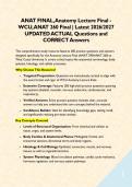
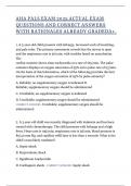
Abdominopelvic Quadrants: abdomen and pelvic regions can be subdivided into 4 regions
o Right upper quadrant (RUQ) liver, gallbladder, right kidney
o Left upper quadrant (LUQ) stomach, pancreas, left kidney, spleen
o Right lower quadrant (RLQ) cecum, appendix
o Lower left quadrant (LLQ) most of small intestine and portions of large intestine
Abdominopelvic Regions: abdomen and pelvic region subdivided into 9 regions
** Anatomical Directions **
Superior/Inferior Medial/ Lateral Proximal/Distal
Anterior/Posterior Deep/Superficial
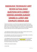
** Sectional Anatomy **
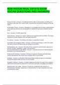
Sagittal Cut: separating left/right
o Midsagittal: separating left/right EQUALLY
o Parasagittal: separating left/right UNEQUALLY
Transverse Cut: separating superior/inferior
Frontal Cut: separating anterior/posterior
Oblique Cut: separating at an angle
, ** Anatomical Regions **
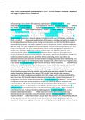
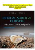
Posterior Cavity: Consists of Cranial cavity (brain) Spinal Cavity (spinal cord)
Anterior Cavity (ventral cavity) Consists of Thoracic, Abdominal, Pelvic Cavity
Membrane nearest the wall of body (farthest from the organ) Parietal membrane
Membrane FARTHEST from the wall of body (nearest the organs) visceral membrane
Pleural cavity: lungs
Pericardial cavity: heart
Mediastinal Cavity: space b/t the apex of the lungs
Peritoneal Cavity stomach, intestines, spleen liver
Pelvic cavity urinary bladder
** QUIZ 1 – CHAPTERS 1-3 **
Cells consists of: (1) Cytoplasm (2) Plasmalemma
Cytoplasm consists of: (1) Cytosol (2) Organelles
Cytosol: fluid within cell
Plasmalemma: 2 layers of fat bound together to make the barrier of the cell
Phagocytosis: Membrane brining solid particles inside of the cell
Cytoskeleton: weaved proteins used to increase surface area of the cell
Ribosomes unites used to assemble proteins
Free Ribosomes: ribosomes attached to the Rough ER
Fixed Ribosomes: ribosomes attached to the Rough ER
Mitochondria: produces ATP using metabolic pathways (O2 + Glucose = ATP)
Nucleus: POWERHOUSE OF THE CELL - creates DNA for the cell
Smooth ER: synthesizes lipids, carbs, steroids, etc
Rough ER: contains ribosomes to create proteins
Golgi Apparatus: synthesizes/ PACKAGE and SHIPS secretions from within the cell
Lysosomes: spherical membrane that secretes enzymes to destroy parts of the cell
Autolysis: rapture of the lysosome to kill the cell
Peroxisomes: spherical membrane that secretes catalase in order to break down hydrogen peroxide
Catalase pathway: gets rid of extra oxygen on H2O to create CO2
Gap Junctions: stations on cells with proteins allowing diffusion from one cell to another
Tight Junction: stations that attach 2 cells to prevent mvt of H2O and other molecules from passing b/t the cells (can be easily torn
apart)
Desmosomes: anchoring junctions that hook cells together to prevent peeling to occur
Epithelial Tissue CELLULARITY: cells bound close together
Epithelial Tissue POLARITY: Apical (outside surface) and basal (facing inside) surfaces
Epithelial Tissue Vascularity: Tissue is avascular (no blood flow)
Microvilli: tiny folds at the top of the cell to increase Surface area ex) intestine to increase absorption
Cilia: hair like projection at the top to transport substances over the cell
ex) found in respiratory tract to move foreign substances out of the way.
Simple epithelium: SINGLE layer of cell
Stratified epithelium: MULTIPLE LAYERS OF CELL
Squamous epithelial cell: large flattened cell with small round nucleus
Cuboidal Cells: cube structure, centered round nucleus
Columnar cell: column structure, nucleus at base
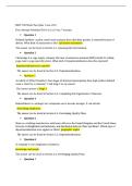
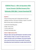
Name Description Function Location
Simple Squamous Epithelium Delicate layer of squamous Lubrication/diffusion Alveoli of lungs
cell
Stratified Squamous Epithelium Multi-layer w/squamous cell Protect against abrasion Skin (keratinized/tough)
@ apical surface Vaginal Canal
(nonkeratinized/moist)
Simple Cuboidal Epithelium Single layer cuboidal cell Secretion/absorption Thyroid gland
Simple Columnar Epithelium Single layer of columnar cell Secretion/absorption Lining of stomach/intestine
Pseudostratified Columnar Fake layers Secretion/absorption Trachea/lungs
Epithelium
Exocrine glands: secretions that travel through ducts to epithelial surface
Endocrine gland: secretions travel through bloodstream as hormones
Anatomy is the study of internal/external structures and the physical relationship b/t body parts.
Physiology: study of human body functions
Histology: study of structure/properties of tissue
Cytology: analyzes the internal structure of cell (smallest unit of life)
Surface anatomy: study of general anatomical form
Regional anatomy: study of superficial/internal features in a specific area of body such as head, neck, or trunk
Systemic anatomy: study of anatomy based upon the body’s organ systems.
Surgical Anatomy: landmarks important for surgical procedures
Radiographic Anatomy: anatomical structures that are visualized by specialized procedures performed on an intact
body
Gross Anatomy: (macroscopic anatomy) is the study of structures & features that are visible to the naked eye
Developmental anatomy: study the changes in form that take place b/t conception and physical maturity
Embryology: study of embryonic development, focusing on the first 2 months after fertilization.
Comparative anatomy: considers the similarities and relationships in anatomic organization of different animals.
Clinical anatomy: anatomical features that undergo characteristic changes during illness
** Levels of Organization (Simple Complex) **
Cell: smallest living unit in the body. Consist of organelles
Tissue: many cells and some surrounding material
Ex) epithelial, muscular, neural, and CT
Organ: combination of tissues that perform complex functions
Ex) heart consist of epithelial, muscular, neural and CT
Organ system: group of organs that function together to produce coordinated effects
Ex) stomach, small intestine, liver, gallbladder, pancreas = digestive system
Organism
**Anatomical Position **
Anatomical position:
o Standing w/feet flat on floor
o Hands are at the side
o Palms are facing forward
Supine: lying down (face UP) in the anatomical position
Prone: lying down (face DOWN) in anatomical position
Abdominopelvic Quadrants: abdomen and pelvic regions can be subdivided into 4 regions
o Right upper quadrant (RUQ) liver, gallbladder, right kidney
o Left upper quadrant (LUQ) stomach, pancreas, left kidney, spleen
o Right lower quadrant (RLQ) cecum, appendix
o Lower left quadrant (LLQ) most of small intestine and portions of large intestine
Abdominopelvic Regions: abdomen and pelvic region subdivided into 9 regions
** Anatomical Directions **
Superior/Inferior Medial/ Lateral Proximal/Distal
Anterior/Posterior Deep/Superficial
** Sectional Anatomy **
Sagittal Cut: separating left/right
o Midsagittal: separating left/right EQUALLY
o Parasagittal: separating left/right UNEQUALLY
Transverse Cut: separating superior/inferior
Frontal Cut: separating anterior/posterior
Oblique Cut: separating at an angle
, ** Anatomical Regions **
Posterior Cavity: Consists of Cranial cavity (brain) Spinal Cavity (spinal cord)
Anterior Cavity (ventral cavity) Consists of Thoracic, Abdominal, Pelvic Cavity
Membrane nearest the wall of body (farthest from the organ) Parietal membrane
Membrane FARTHEST from the wall of body (nearest the organs) visceral membrane
Pleural cavity: lungs
Pericardial cavity: heart
Mediastinal Cavity: space b/t the apex of the lungs
Peritoneal Cavity stomach, intestines, spleen liver
Pelvic cavity urinary bladder
** QUIZ 1 – CHAPTERS 1-3 **
Cells consists of: (1) Cytoplasm (2) Plasmalemma
Cytoplasm consists of: (1) Cytosol (2) Organelles
Cytosol: fluid within cell
Plasmalemma: 2 layers of fat bound together to make the barrier of the cell
Phagocytosis: Membrane brining solid particles inside of the cell
Cytoskeleton: weaved proteins used to increase surface area of the cell
Ribosomes unites used to assemble proteins
Free Ribosomes: ribosomes attached to the Rough ER
Fixed Ribosomes: ribosomes attached to the Rough ER
Mitochondria: produces ATP using metabolic pathways (O2 + Glucose = ATP)
Nucleus: POWERHOUSE OF THE CELL - creates DNA for the cell
Smooth ER: synthesizes lipids, carbs, steroids, etc
Rough ER: contains ribosomes to create proteins
Golgi Apparatus: synthesizes/ PACKAGE and SHIPS secretions from within the cell
Lysosomes: spherical membrane that secretes enzymes to destroy parts of the cell
Autolysis: rapture of the lysosome to kill the cell
Peroxisomes: spherical membrane that secretes catalase in order to break down hydrogen peroxide
Catalase pathway: gets rid of extra oxygen on H2O to create CO2
Gap Junctions: stations on cells with proteins allowing diffusion from one cell to another
Tight Junction: stations that attach 2 cells to prevent mvt of H2O and other molecules from passing b/t the cells (can be easily torn
apart)
Desmosomes: anchoring junctions that hook cells together to prevent peeling to occur
Epithelial Tissue CELLULARITY: cells bound close together
Epithelial Tissue POLARITY: Apical (outside surface) and basal (facing inside) surfaces
Epithelial Tissue Vascularity: Tissue is avascular (no blood flow)
Microvilli: tiny folds at the top of the cell to increase Surface area ex) intestine to increase absorption
Cilia: hair like projection at the top to transport substances over the cell
ex) found in respiratory tract to move foreign substances out of the way.
Simple epithelium: SINGLE layer of cell
Stratified epithelium: MULTIPLE LAYERS OF CELL
Squamous epithelial cell: large flattened cell with small round nucleus
Cuboidal Cells: cube structure, centered round nucleus
Columnar cell: column structure, nucleus at base
Name Description Function Location
Simple Squamous Epithelium Delicate layer of squamous Lubrication/diffusion Alveoli of lungs
cell
Stratified Squamous Epithelium Multi-layer w/squamous cell Protect against abrasion Skin (keratinized/tough)
@ apical surface Vaginal Canal
(nonkeratinized/moist)
Simple Cuboidal Epithelium Single layer cuboidal cell Secretion/absorption Thyroid gland
Simple Columnar Epithelium Single layer of columnar cell Secretion/absorption Lining of stomach/intestine
Pseudostratified Columnar Fake layers Secretion/absorption Trachea/lungs
Epithelium
Exocrine glands: secretions that travel through ducts to epithelial surface
Endocrine gland: secretions travel through bloodstream as hormones