
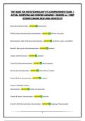
Alcian Blue demonstrates - ANSWER Acid mucins
ATPase Enyme Histochemistry demonstrates - ANSWER ATPase in muscles
Bielschowsky's Silver Technique demonatrates - ANSWER Dendrites, axons, neurofibrils
Brown & Brenn gram stain demonstrates - ANSWER bacteria
Congo red demonstrates - ANSWER amyloid
Cresyl Echt Violet demonstrates - ANSWER Nissl substance
Geimsa stain demonstrates - ANSWER mast cells or H. pylori
Gomori Burtner demonstrates - ANSWER melanin
Gomori's Aldehyde Fuchsin
demonstrates - ANSWER elastic and mast cells
Gordon & Sweet's demonstrates - ANSWER reticulin
Grocott's Methenamine Silver demonstrates - ANSWER fungi (eg. Pneumocystis)
1
,Hematoxylin & Eosin shows - ANSWER general stain
Immunohistotochemistry - ANSWER Anything an antibody can be made for
Luxol fast blue stain shows - ANSWER myelin
Toluidine blue stain shows - ANSWER mast cells or H. pylory
Masson's Trichrome shows - ANSWER collagen
Oil Red O technique shows - ANSWER neutral fats (simple fats)
Papanicolaou technique shows - ANSWER cytology smears
Periodic Acid Schiff's shows - ANSWER neutral mucin, glycogen
Perl's Prussian Blue shows - ANSWER Hemosiderin
Southgate's mucicarmine - ANSWER epithelial acid mucins
Verhoeff's Van Gieson - ANSWER elastic
Van Kossa shows - ANSWER calcium
Ziehl Neelsen shows - ANSWER Acid fast bacilli (T.B.)
2
,Nuclei in Gomori's Aldehyde Fuchsin technique - ANSWER yellow - no specific stain for
nuclei
Mast cells in a Gomori's Aldehyde Fuchsin technique - ANSWER purple - GAF not specific
for elastic
Gram positive bacteria if over-differentiated after crystal violet - ANSWER red
Nuclei in the Brown and Brenn Gram stain if over differentiated with picric acid/acetone -
ANSWER yellow
Fungi in the Periodic Acid Schiff (PAS) technique - ANSWER magenta - chitin binds with
Schiffs
Fungi in the Grocott's Methenamine Silver technique - ANSWER black - silver impregna-
tion
Neutral mucins in a PAS technique if potassium permanganate is used as the oxidizing agent
- ANSWER light pink - colour of background - CHO overoxidized from aldehydes to acid
state
Cytoplasm in an H&E if over differentiated with acid alcohol - ANSWER pink - only affects
nuclei
Epithelial acid mucins in the Southgate's mucicarmine technique - ANSWER red
Cytoplasm in an H&E if the blueing agent is not properly removed - ANSWER very pale
pink
Nuclei in the Congo Red technique - ANSWER blue - stained with aluminum hematoxylin
3
, Calcium in the Von Kossa technique - ANSWER black - silver impregnation
Glycogen, after diastase in the Periodic Acid Schiff technique - ANSWER pale pink - back-
ground colour
Mycobacterium after Ziehl Neelsen technique - ANSWER red - AFB stain with carbol fuch-
sin
Mycobacterium after Ziehl Neelsen technique - ANSWER red - AFB stain with carbol fuch-
sin
Streptococcus after Ziehl Neelsen technique - ANSWER blue - colour of background
Neutral lipids for Oil Red O cut on a cryostat - ANSWER red to orange
Neutral lipids for Oil Red O cut from wax block - ANSWER not demonstrated - removed
Hemosiderin after a Perl's Prussian Blue - ANSWER blue
Nuclei after Masson's trichrome using an aluminum Hematoxylin - ANSWER red - same as
cytoplasm
Fine elastic fibres in Verhoeff's Van Gieson (VVG) after leaving in Van Gieson reagent too long
- ANSWER not black/not demonstrated - iron hematoxylin removed by the acid
Cytoplasm if underdifferentiated in the VVG technique - ANSWER grey to black
4
ATPase Enyme Histochemistry demonstrates - ANSWER ATPase in muscles
Bielschowsky's Silver Technique demonatrates - ANSWER Dendrites, axons, neurofibrils
Brown & Brenn gram stain demonstrates - ANSWER bacteria
Congo red demonstrates - ANSWER amyloid
Cresyl Echt Violet demonstrates - ANSWER Nissl substance
Geimsa stain demonstrates - ANSWER mast cells or H. pylori
Gomori Burtner demonstrates - ANSWER melanin
Gomori's Aldehyde Fuchsin
demonstrates - ANSWER elastic and mast cells
Gordon & Sweet's demonstrates - ANSWER reticulin
Grocott's Methenamine Silver demonstrates - ANSWER fungi (eg. Pneumocystis)
1
,Hematoxylin & Eosin shows - ANSWER general stain
Immunohistotochemistry - ANSWER Anything an antibody can be made for
Luxol fast blue stain shows - ANSWER myelin
Toluidine blue stain shows - ANSWER mast cells or H. pylory
Masson's Trichrome shows - ANSWER collagen
Oil Red O technique shows - ANSWER neutral fats (simple fats)
Papanicolaou technique shows - ANSWER cytology smears
Periodic Acid Schiff's shows - ANSWER neutral mucin, glycogen
Perl's Prussian Blue shows - ANSWER Hemosiderin
Southgate's mucicarmine - ANSWER epithelial acid mucins
Verhoeff's Van Gieson - ANSWER elastic
Van Kossa shows - ANSWER calcium
Ziehl Neelsen shows - ANSWER Acid fast bacilli (T.B.)
2
,Nuclei in Gomori's Aldehyde Fuchsin technique - ANSWER yellow - no specific stain for
nuclei
Mast cells in a Gomori's Aldehyde Fuchsin technique - ANSWER purple - GAF not specific
for elastic
Gram positive bacteria if over-differentiated after crystal violet - ANSWER red
Nuclei in the Brown and Brenn Gram stain if over differentiated with picric acid/acetone -
ANSWER yellow
Fungi in the Periodic Acid Schiff (PAS) technique - ANSWER magenta - chitin binds with
Schiffs
Fungi in the Grocott's Methenamine Silver technique - ANSWER black - silver impregna-
tion
Neutral mucins in a PAS technique if potassium permanganate is used as the oxidizing agent
- ANSWER light pink - colour of background - CHO overoxidized from aldehydes to acid
state
Cytoplasm in an H&E if over differentiated with acid alcohol - ANSWER pink - only affects
nuclei
Epithelial acid mucins in the Southgate's mucicarmine technique - ANSWER red
Cytoplasm in an H&E if the blueing agent is not properly removed - ANSWER very pale
pink
Nuclei in the Congo Red technique - ANSWER blue - stained with aluminum hematoxylin
3
, Calcium in the Von Kossa technique - ANSWER black - silver impregnation
Glycogen, after diastase in the Periodic Acid Schiff technique - ANSWER pale pink - back-
ground colour
Mycobacterium after Ziehl Neelsen technique - ANSWER red - AFB stain with carbol fuch-
sin
Mycobacterium after Ziehl Neelsen technique - ANSWER red - AFB stain with carbol fuch-
sin
Streptococcus after Ziehl Neelsen technique - ANSWER blue - colour of background
Neutral lipids for Oil Red O cut on a cryostat - ANSWER red to orange
Neutral lipids for Oil Red O cut from wax block - ANSWER not demonstrated - removed
Hemosiderin after a Perl's Prussian Blue - ANSWER blue
Nuclei after Masson's trichrome using an aluminum Hematoxylin - ANSWER red - same as
cytoplasm
Fine elastic fibres in Verhoeff's Van Gieson (VVG) after leaving in Van Gieson reagent too long
- ANSWER not black/not demonstrated - iron hematoxylin removed by the acid
Cytoplasm if underdifferentiated in the VVG technique - ANSWER grey to black
4


