Bio 1106 Unit 3
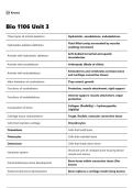
Three types of animal skeletons Hydrostatic, exoskeletons, endoskeletons
Fluid‑filled cavity surrounded by muscles
Hydrostatic skeleton definition
enabling movement
Soft‑bodied terrestrial and aquatic
Animals with hydrostatic skeletons
invertebrates
Animals with exoskeletons Arthropods (Made of chitin)
Echinoderms and vertebrates (contains bone
Animals with endoskeletons
and cartilage-connective tissue)
Main limitation of exoskeletons They restrict growth
Functions of exoskeletons Protection, muscle attachment, rigid support
Internal support, muscle attachment, organ
Functions of endoskeletons
protection
Collagen (flexibility) + hydroxyapatite
Composition of bone
(rigidity)
Cartilage tissue characteristics Tough, flexible, avascular connective tissue
Cells that maintain cartilage Chondrocytes
Osteoblasts Cells that build bone
Osteocytes Cells that maintain bone
Osteoclasts Cells that break down bone
Structural unit of compact bone housing blood
Haversian system
vessels and nerves
Bone forms within connective tissue (flat
Intramembranous bone development
bones)
Endochondral bone development Bone replaces a cartilage model (long bones)
, Balance of osteoblasts (build) and osteoclasts
Bone remodeling
(break down)
Smooth muscle Involuntary, internal organs
Cardiac muscle Involuntary, contracts as a unit, heart
Skeletal muscle Voluntary, attached to bones
Smallest functional unit of muscle
Sarcomere
contraction
Bundles of myofilaments responsible for
Myofibrils
contraction
Striations Alternating dark/light bands in skeletal muscle
ATP binding to myosin Causes myosin to release actin
ATP → ADP + Pi Cocks the myosin head
Myosin binds actin Cross‑bridge formation
Power stroke Myosin pulls actin toward sarcomere center
New ATP binding Myosin detaches from actin
Rigor mortis cause No ATP to detach myosin from actin
Low Ca²⁺ Tropomyosin blocks actin binding sites
Ca²⁺ binds troponin, moves tropomyosin,
High Ca²⁺
allows contraction
Actin and myosin slide; filaments do not
Sliding filament theory
shorten
Acetylcholine (ACh) Neurotransmitter at NMJ
Motor end plate Muscle membrane region with ACh receptors
Na⁺ influx Causes muscle depolarization
T‑tubules Carry depolarization deep into muscle fiber
Sarcoplasmic reticulum Stores and releases Ca²⁺
One motor neuron + all muscle fibers it
Motor unit
innervates
, Recruitment Activating more motor units to increase force
Muscle twitch Single contraction‑relaxation event
Summation Multiple twitches combine to increase force
Sustained, powerful contraction with partial
Incomplete tetanus
relaxation
Complete tetanus Maximum tension, no relaxation
Aerobic, many mitochondria, high
Slow‑twitch fibers
myoglobin, fatigue‑resistant
Anaerobic, fewer mitochondria, low myoglobin,
Fast‑twitch fibers
fatigue quickly
Unit of energy; amount needed to raise 1 kg
Calorie (kilocalorie)
of water by 1°C
Basal metabolic rate (BMR) Minimum energy required at rest
Provide amino acids for building tissues and
Proteins
enzymes
Essential amino acids Amino acids that must be obtained from diet
Number of amino acids required by humans 11 of the 20 must come from diet
Tubular digestive pathway from mouth to
GI tract
anus
Accessory organs Liver, pancreas, gallbladder, salivary glands
Innermost layer responsible for secretion,
Mucosa
absorption, protection
Stratified squamous (mouth, esophagus,
Epithelium of mucosa anus) or simple columnar (stomach,
intestines)
Connective tissue with blood vessels,
Lamina propria
lymphatics, immune cells
Muscularis mucosae Thin smooth muscle increasing surface area
Dense connective tissue with vessels, glands,
Submucosa
and nerve plexus
Three types of animal skeletons Hydrostatic, exoskeletons, endoskeletons
Fluid‑filled cavity surrounded by muscles
Hydrostatic skeleton definition
enabling movement
Soft‑bodied terrestrial and aquatic
Animals with hydrostatic skeletons
invertebrates
Animals with exoskeletons Arthropods (Made of chitin)
Echinoderms and vertebrates (contains bone
Animals with endoskeletons
and cartilage-connective tissue)
Main limitation of exoskeletons They restrict growth
Functions of exoskeletons Protection, muscle attachment, rigid support
Internal support, muscle attachment, organ
Functions of endoskeletons
protection
Collagen (flexibility) + hydroxyapatite
Composition of bone
(rigidity)
Cartilage tissue characteristics Tough, flexible, avascular connective tissue
Cells that maintain cartilage Chondrocytes
Osteoblasts Cells that build bone
Osteocytes Cells that maintain bone
Osteoclasts Cells that break down bone
Structural unit of compact bone housing blood
Haversian system
vessels and nerves
Bone forms within connective tissue (flat
Intramembranous bone development
bones)
Endochondral bone development Bone replaces a cartilage model (long bones)
, Balance of osteoblasts (build) and osteoclasts
Bone remodeling
(break down)
Smooth muscle Involuntary, internal organs
Cardiac muscle Involuntary, contracts as a unit, heart
Skeletal muscle Voluntary, attached to bones
Smallest functional unit of muscle
Sarcomere
contraction
Bundles of myofilaments responsible for
Myofibrils
contraction
Striations Alternating dark/light bands in skeletal muscle
ATP binding to myosin Causes myosin to release actin
ATP → ADP + Pi Cocks the myosin head
Myosin binds actin Cross‑bridge formation
Power stroke Myosin pulls actin toward sarcomere center
New ATP binding Myosin detaches from actin
Rigor mortis cause No ATP to detach myosin from actin
Low Ca²⁺ Tropomyosin blocks actin binding sites
Ca²⁺ binds troponin, moves tropomyosin,
High Ca²⁺
allows contraction
Actin and myosin slide; filaments do not
Sliding filament theory
shorten
Acetylcholine (ACh) Neurotransmitter at NMJ
Motor end plate Muscle membrane region with ACh receptors
Na⁺ influx Causes muscle depolarization
T‑tubules Carry depolarization deep into muscle fiber
Sarcoplasmic reticulum Stores and releases Ca²⁺
One motor neuron + all muscle fibers it
Motor unit
innervates
, Recruitment Activating more motor units to increase force
Muscle twitch Single contraction‑relaxation event
Summation Multiple twitches combine to increase force
Sustained, powerful contraction with partial
Incomplete tetanus
relaxation
Complete tetanus Maximum tension, no relaxation
Aerobic, many mitochondria, high
Slow‑twitch fibers
myoglobin, fatigue‑resistant
Anaerobic, fewer mitochondria, low myoglobin,
Fast‑twitch fibers
fatigue quickly
Unit of energy; amount needed to raise 1 kg
Calorie (kilocalorie)
of water by 1°C
Basal metabolic rate (BMR) Minimum energy required at rest
Provide amino acids for building tissues and
Proteins
enzymes
Essential amino acids Amino acids that must be obtained from diet
Number of amino acids required by humans 11 of the 20 must come from diet
Tubular digestive pathway from mouth to
GI tract
anus
Accessory organs Liver, pancreas, gallbladder, salivary glands
Innermost layer responsible for secretion,
Mucosa
absorption, protection
Stratified squamous (mouth, esophagus,
Epithelium of mucosa anus) or simple columnar (stomach,
intestines)
Connective tissue with blood vessels,
Lamina propria
lymphatics, immune cells
Muscularis mucosae Thin smooth muscle increasing surface area
Dense connective tissue with vessels, glands,
Submucosa
and nerve plexus