
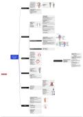
🔹 The larynx is the voice box
formed of cartilages connected
to each other by ligaments ,
moved by muscles and lined by Laryngeal Cavity
🔹
mucous membrane
Situated in midline of neck
extend from upper border of
Supraglottic area
Glottic area
Subglottic area
epiglottis to lower border of
cricoid cartilage ( from C3 to
lower border of C6 )
Defination of larynx
🔹
EPITHELIUM OF LARYNX
Respiratory epithelium ( pseudostratified cilliated
columnar epithelium )
All larynx except , vocal cord , epiglottis
Stratified squamous epithelium :
Site of mechanical stress or abrasion
1- vocal cord
2- epiglottis
🔷3 single cartilage
🔹 Thyroid cartilage:
Largest , It resembles open book
Formed of 2 lamina ( alae ) fused anterior at an angle ( 90 – 110 )
deficint superior forming thyroid notch
Thyroid angle more prominant in male ( adam’s apple )
🔹
Each lamina has superior and inferior thyroid horns
Cricoid cartilage:
stronge , signet ring in appearance
🔹
Wide posterior lamina , narrow anterior arch
3 single and 3 paired epiglottis:
Fibroelastic , leaf like in appearance
🔹
cartilages:
Single cartilage :
Thyroid cartilage
Its lower part attached to the mid-point of posterior surface of
thyroid angle ( petiol )
It has multible pits on its surface caused by the seromucinous glands
Cricoid cartilage
🔹
Epiglottis
🔷
🔹 Paired cartilages
Paired cartilages :
Arytenoid cartilages Arytenoid cartilages :
Corniculate cartilages Pyramidal in shape , it has apex directed upwards and base
Cuniform cartilages articulating with the upper surface of posterior part of cricoid (
lamina )
It has vocal process directed anteriorly for attachement of vocal
fold
And muscular process directed laterally for muscle attachment (
🔹
posterior And lateral cricoarytenoid muscle )
Corniculate cartilages :
Articulate with the apex of arytenoid
🔹 Cuniform cartilages :
Present on the aryepiglottic fold
cartilages and ligaments 🔹 Extrinsic membrane :
1-Thyrohyoid : between hyoid bone and thyroid cartilage
2-Cricothyroid : between cricoid and thyroid cartilage
3-Cricotraceal : between cricoid and trachea
Hyo
4-epiglottic : between hyoid bone and epiglottis
5-Thyro-epiglottic : between thyroid cartilage and epiglottis
Ligaments and membranes
🔹 Intrinsic membrane :
1- Quadrangular membrane : upper part of larynx , extend from
epiglottis to the arytenoid cartilage , supports the false vocal cords .
2- Conus elasticus ( Cricothyroid membrane ) : lower part of
larynx , connects the cricoid and thyroid cartilage , supports the
true vocal cord .
Laryngeal inlet boundaries :
Anterior --- epiglottis
Laryngeal inlet Posterior --- arytenoid
Laterally --- ary-epiglottic fold
🔹 1-True vocal folds :
attached anterior to the
midpoint of the post. Surface
of thyroid angle and post. To 🔷
🔹 Protective function:
1- Reflex closure during swallowing by 3
🔹
the vocal process of arytenoid
2-False vocal folds :
above the level of the true
mechanism
- Ary-epiglottic folds
Anatomy and 🔹
vocal cord - False vocal folds
Physiology of
3-Aryepiglottic folds :
attached anterior to epiglottis 🔹
🔹
- True vocal folds
2- Reflex elevation of larynx during swallowing
3- Reflex inhibition of respiratory during
and posterior to arytenoid
LARYNX Laryngeal folds 🔹
swallowing
4- Reflex cough ( FB )
🔹
Position of vocal cord
Median : the V.C is in Function of the larynx : 🔷 2- Respiratory function :
🔹
midline position
Paramedian : the V.C in
Physiology of the larynx
1- Protective function
2- Respiratory function
The vocal folds become fully abducted during
🔹
paramedian position inspiration
3- Phonatory function
🔹
Cadaveric : the V.C is in
intermediate position
Abduction : the V.C is in
4- Fixation of chest
🔷 3- Phonatory function :
The vocal folds become adducted during
🔹
abduction position
Full abduction : the V.C is
in full abduction position
phonation during expiration
4. Fixation of chest :
The vocal fold become adducted to increase intra
thoracic pressure during lifting heavy weights ,
labour and defecation .
🔷 Pre-epiglottic space :
Infront of epiglottis
🔹
Boundaries :
🔹 Anterior : thyrohyoid membrane
🔹Posterior : epiglottis
Superior : hyo-epiglottic ligament
Space related to the larynx
by fatema okoff 🔷 Paraglottic space
Present to the side of the glottis
🔹
Boundaries
🔹Medialy : true and false vocal cord
Laterally : thyroid cartilage
N.B : if carcinoma reach to the paraglottic space lead to vocal
fold fixation
🔹
Extrinsic Muscles :
Suprahyoid group muscle
Digastric muscle
Stylohyoid muscle
Mylohyoid muscle
Geniohyoid muscle
🔹
Stylopharyngeus muscle
Infrahyoid group muscles
Sternohyoid muscle
🔷
Omohyoid muscle
Extrinsic Muscles : Sternothyroid
Suprahyoid group muscles Thyrohyoid
🔷
Infrahyoid group muscles
Muscles of larynx Intrinsic Muscles :
Abductors muscle
Adductors muscles
Tensor muscle
🔹
Intrinsic Muscles
Adductors muscle :
Lateral Cricoarytenoid muscle
Thyroarytenoid muscle
🔹
Inter arytenoid muscle
Abductors muscle :
Posterior Cricoarytenoid muscle
Tensors muscle :
Cricothyroid muscle
Vocalis muscle
Innervation of the larynx
🔷
🔹 Sensory Innervation :
Superior laryngeal nerve ( internal laryngeal branche ) :
🔹Recurrent laryngeal nerve : below the V.C
above the V.C
🔷
🔹 Motor Innervation :
Recurrent laryngeal nerve : all larynx except cricothyroid
🔹
muscle
Superior laryngeal nerve ( external laryngeal branche) :
Cricothyroid muscle
supply 🔹
Blood supply
Superior laryngeal artery
( from superior thyroid artery )
🔹( from
Inferior laryngeal artery
inferior thyroid artery )
Lymphatic drainage
🔹upper
Supragluttic area :
🔹lower deep cervical LN
Subglottic area :
🔹noGlottic
deep cervical LN
area :
lymphatic drainage
formed of cartilages connected
to each other by ligaments ,
moved by muscles and lined by Laryngeal Cavity
🔹
mucous membrane
Situated in midline of neck
extend from upper border of
Supraglottic area
Glottic area
Subglottic area
epiglottis to lower border of
cricoid cartilage ( from C3 to
lower border of C6 )
Defination of larynx
🔹
EPITHELIUM OF LARYNX
Respiratory epithelium ( pseudostratified cilliated
columnar epithelium )
All larynx except , vocal cord , epiglottis
Stratified squamous epithelium :
Site of mechanical stress or abrasion
1- vocal cord
2- epiglottis
🔷3 single cartilage
🔹 Thyroid cartilage:
Largest , It resembles open book
Formed of 2 lamina ( alae ) fused anterior at an angle ( 90 – 110 )
deficint superior forming thyroid notch
Thyroid angle more prominant in male ( adam’s apple )
🔹
Each lamina has superior and inferior thyroid horns
Cricoid cartilage:
stronge , signet ring in appearance
🔹
Wide posterior lamina , narrow anterior arch
3 single and 3 paired epiglottis:
Fibroelastic , leaf like in appearance
🔹
cartilages:
Single cartilage :
Thyroid cartilage
Its lower part attached to the mid-point of posterior surface of
thyroid angle ( petiol )
It has multible pits on its surface caused by the seromucinous glands
Cricoid cartilage
🔹
Epiglottis
🔷
🔹 Paired cartilages
Paired cartilages :
Arytenoid cartilages Arytenoid cartilages :
Corniculate cartilages Pyramidal in shape , it has apex directed upwards and base
Cuniform cartilages articulating with the upper surface of posterior part of cricoid (
lamina )
It has vocal process directed anteriorly for attachement of vocal
fold
And muscular process directed laterally for muscle attachment (
🔹
posterior And lateral cricoarytenoid muscle )
Corniculate cartilages :
Articulate with the apex of arytenoid
🔹 Cuniform cartilages :
Present on the aryepiglottic fold
cartilages and ligaments 🔹 Extrinsic membrane :
1-Thyrohyoid : between hyoid bone and thyroid cartilage
2-Cricothyroid : between cricoid and thyroid cartilage
3-Cricotraceal : between cricoid and trachea
Hyo
4-epiglottic : between hyoid bone and epiglottis
5-Thyro-epiglottic : between thyroid cartilage and epiglottis
Ligaments and membranes
🔹 Intrinsic membrane :
1- Quadrangular membrane : upper part of larynx , extend from
epiglottis to the arytenoid cartilage , supports the false vocal cords .
2- Conus elasticus ( Cricothyroid membrane ) : lower part of
larynx , connects the cricoid and thyroid cartilage , supports the
true vocal cord .
Laryngeal inlet boundaries :
Anterior --- epiglottis
Laryngeal inlet Posterior --- arytenoid
Laterally --- ary-epiglottic fold
🔹 1-True vocal folds :
attached anterior to the
midpoint of the post. Surface
of thyroid angle and post. To 🔷
🔹 Protective function:
1- Reflex closure during swallowing by 3
🔹
the vocal process of arytenoid
2-False vocal folds :
above the level of the true
mechanism
- Ary-epiglottic folds
Anatomy and 🔹
vocal cord - False vocal folds
Physiology of
3-Aryepiglottic folds :
attached anterior to epiglottis 🔹
🔹
- True vocal folds
2- Reflex elevation of larynx during swallowing
3- Reflex inhibition of respiratory during
and posterior to arytenoid
LARYNX Laryngeal folds 🔹
swallowing
4- Reflex cough ( FB )
🔹
Position of vocal cord
Median : the V.C is in Function of the larynx : 🔷 2- Respiratory function :
🔹
midline position
Paramedian : the V.C in
Physiology of the larynx
1- Protective function
2- Respiratory function
The vocal folds become fully abducted during
🔹
paramedian position inspiration
3- Phonatory function
🔹
Cadaveric : the V.C is in
intermediate position
Abduction : the V.C is in
4- Fixation of chest
🔷 3- Phonatory function :
The vocal folds become adducted during
🔹
abduction position
Full abduction : the V.C is
in full abduction position
phonation during expiration
4. Fixation of chest :
The vocal fold become adducted to increase intra
thoracic pressure during lifting heavy weights ,
labour and defecation .
🔷 Pre-epiglottic space :
Infront of epiglottis
🔹
Boundaries :
🔹 Anterior : thyrohyoid membrane
🔹Posterior : epiglottis
Superior : hyo-epiglottic ligament
Space related to the larynx
by fatema okoff 🔷 Paraglottic space
Present to the side of the glottis
🔹
Boundaries
🔹Medialy : true and false vocal cord
Laterally : thyroid cartilage
N.B : if carcinoma reach to the paraglottic space lead to vocal
fold fixation
🔹
Extrinsic Muscles :
Suprahyoid group muscle
Digastric muscle
Stylohyoid muscle
Mylohyoid muscle
Geniohyoid muscle
🔹
Stylopharyngeus muscle
Infrahyoid group muscles
Sternohyoid muscle
🔷
Omohyoid muscle
Extrinsic Muscles : Sternothyroid
Suprahyoid group muscles Thyrohyoid
🔷
Infrahyoid group muscles
Muscles of larynx Intrinsic Muscles :
Abductors muscle
Adductors muscles
Tensor muscle
🔹
Intrinsic Muscles
Adductors muscle :
Lateral Cricoarytenoid muscle
Thyroarytenoid muscle
🔹
Inter arytenoid muscle
Abductors muscle :
Posterior Cricoarytenoid muscle
Tensors muscle :
Cricothyroid muscle
Vocalis muscle
Innervation of the larynx
🔷
🔹 Sensory Innervation :
Superior laryngeal nerve ( internal laryngeal branche ) :
🔹Recurrent laryngeal nerve : below the V.C
above the V.C
🔷
🔹 Motor Innervation :
Recurrent laryngeal nerve : all larynx except cricothyroid
🔹
muscle
Superior laryngeal nerve ( external laryngeal branche) :
Cricothyroid muscle
supply 🔹
Blood supply
Superior laryngeal artery
( from superior thyroid artery )
🔹( from
Inferior laryngeal artery
inferior thyroid artery )
Lymphatic drainage
🔹upper
Supragluttic area :
🔹lower deep cervical LN
Subglottic area :
🔹noGlottic
deep cervical LN
area :
lymphatic drainage


