

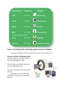
Lecture 1 The Resting State: physiology, cognition and your databases
- According to Gazzaniga, the field of “cognitive neuroscience” is only ~40 years old
Positron emission tomography (PET)
Principle: Radioactive material is injected
(e.g., fluorodeoxyglucose, FDG).
The FDG sugar accumulates in brain areas
that are metabolically active.
The decay of FDG gives off a neutron and a
positron
=> the positron hits an electron and
annihilates into 2 gamma rays, which are
detected.
,…metabolic imaging with PET has poor temporal resolution (~10-20 s)
PET is an important functional imaging technique that has been widely used since
the 1980s, especially in drug research and neuroscience.
In PET, a radioactive tracer (such as fluorine, oxygen, carbon, or nitrogen) is injected
or inhaled. This tracer travels through the bloodstream to active brain areas (those
with high oxygen or glucose consumption). The most commonly used tracer is FDG
(fluorodeoxyglucose), a sugar.
As the tracer decays, it emits a positron that collides with an electron. This results in
annihilation and the emission of two gamma rays, which are detected by the
scanner. This allows for the creation of a functional image of brain activity.
● Temporal resolution: 10–20 seconds
● Spatial resolution: approximately 5 mm
PET enables the measurement of physiological processes, such as glucose
metabolism, perfusion, or receptor distribution. Tracers are created by linking
a radionuclide to a biologically active molecule. If this molecule binds to a
neuroreceptor, PET can visualize the location and activity of that receptor.
Examples of applications:
● Raclopride for the dopamine D2 receptor (used in Parkinson’s research)
● Tracers for amyloid plaques (used in Alzheimer’s diagnosis and therapy
monitoring)
● Tracers for serotonin receptors (used in depression studies)
Hoge energiebehoefte van het brein
● De hersenen gebruiken ~20% van de totale energie van het lichaam, zelfs in
rust.
● Per gram gebruikt de hersenen meer energie dan het kloppende hart.
● Toch is de toename in metabolisme tijdens een taak klein (~<5%) → daarom
is het moeilijk om hersenactiviteit direct tijdens één taak te meten.
,De oplossing: subtractiemethode
● Om hersenactiviteit toch goed te meten, gebruiken onderzoekers taakblokken.
Ze vergelijken het PET-signaal (glucoseverbruik) tijdens twee verschillende
taken:
○ Bijv. ‘woord lezen’ – ‘visuele fixatie’
● Zo wordt het verschil in hersenactiviteit zichtbaar, doordat alle algemene
activatie (zoals visuele input) wordt weggefilterd.
Interpretatie van de ‘difference images’ (rechterkolom in figuur)
● Deze beelden tonen waar het brein méér
energie verbruikt bij een specifieke taak
vergeleken met de basistaak.
● De kleurenschaal laat zien hoeveel procent
verschil er is:
○ Geel/rood/wit = grootste toename
in activiteit (tot 5%)
○ Blauw/paars = weinig of geen
verschil
● Voorbeeld:
○ Bij ‘generating verbs’ is vooral de linker frontale cortex actiever dan bij
‘reading words’ → dit wijst op taal productieprocessen.
Early successes of cognitive neuroimaging
Speaking words activates Wernicke’s area, whereas thinking about words activates Broca’s
area in the frontal lobe
, fMRI has largely replaced PET in Cognitive Neuroscience
- Note, fMRI is an
indirect measure
of brain activity!
Uit onderzoek blijkt dat
95% van je hersenen
activiteit laat zien bij
kleine taakjes. Er bleek
dat bepaalde functies
gelokaliseerd zijn in
bepaalde
hersengebieden. Maar
daarnaast veel meer
hersengebieden mee
dan de lokale
functiekaart aantoont.
- According to Gazzaniga, the field of “cognitive neuroscience” is only ~40 years old
Positron emission tomography (PET)
Principle: Radioactive material is injected
(e.g., fluorodeoxyglucose, FDG).
The FDG sugar accumulates in brain areas
that are metabolically active.
The decay of FDG gives off a neutron and a
positron
=> the positron hits an electron and
annihilates into 2 gamma rays, which are
detected.
,…metabolic imaging with PET has poor temporal resolution (~10-20 s)
PET is an important functional imaging technique that has been widely used since
the 1980s, especially in drug research and neuroscience.
In PET, a radioactive tracer (such as fluorine, oxygen, carbon, or nitrogen) is injected
or inhaled. This tracer travels through the bloodstream to active brain areas (those
with high oxygen or glucose consumption). The most commonly used tracer is FDG
(fluorodeoxyglucose), a sugar.
As the tracer decays, it emits a positron that collides with an electron. This results in
annihilation and the emission of two gamma rays, which are detected by the
scanner. This allows for the creation of a functional image of brain activity.
● Temporal resolution: 10–20 seconds
● Spatial resolution: approximately 5 mm
PET enables the measurement of physiological processes, such as glucose
metabolism, perfusion, or receptor distribution. Tracers are created by linking
a radionuclide to a biologically active molecule. If this molecule binds to a
neuroreceptor, PET can visualize the location and activity of that receptor.
Examples of applications:
● Raclopride for the dopamine D2 receptor (used in Parkinson’s research)
● Tracers for amyloid plaques (used in Alzheimer’s diagnosis and therapy
monitoring)
● Tracers for serotonin receptors (used in depression studies)
Hoge energiebehoefte van het brein
● De hersenen gebruiken ~20% van de totale energie van het lichaam, zelfs in
rust.
● Per gram gebruikt de hersenen meer energie dan het kloppende hart.
● Toch is de toename in metabolisme tijdens een taak klein (~<5%) → daarom
is het moeilijk om hersenactiviteit direct tijdens één taak te meten.
,De oplossing: subtractiemethode
● Om hersenactiviteit toch goed te meten, gebruiken onderzoekers taakblokken.
Ze vergelijken het PET-signaal (glucoseverbruik) tijdens twee verschillende
taken:
○ Bijv. ‘woord lezen’ – ‘visuele fixatie’
● Zo wordt het verschil in hersenactiviteit zichtbaar, doordat alle algemene
activatie (zoals visuele input) wordt weggefilterd.
Interpretatie van de ‘difference images’ (rechterkolom in figuur)
● Deze beelden tonen waar het brein méér
energie verbruikt bij een specifieke taak
vergeleken met de basistaak.
● De kleurenschaal laat zien hoeveel procent
verschil er is:
○ Geel/rood/wit = grootste toename
in activiteit (tot 5%)
○ Blauw/paars = weinig of geen
verschil
● Voorbeeld:
○ Bij ‘generating verbs’ is vooral de linker frontale cortex actiever dan bij
‘reading words’ → dit wijst op taal productieprocessen.
Early successes of cognitive neuroimaging
Speaking words activates Wernicke’s area, whereas thinking about words activates Broca’s
area in the frontal lobe
, fMRI has largely replaced PET in Cognitive Neuroscience
- Note, fMRI is an
indirect measure
of brain activity!
Uit onderzoek blijkt dat
95% van je hersenen
activiteit laat zien bij
kleine taakjes. Er bleek
dat bepaalde functies
gelokaliseerd zijn in
bepaalde
hersengebieden. Maar
daarnaast veel meer
hersengebieden mee
dan de lokale
functiekaart aantoont.


