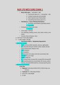
NUR 170 MED SURG EXAM 1
• Body is 60% water o Intracellular – 66%
Fluid volume about 25 L o Extracellular – 33%
Includes intravascular and interstitial
Most important area of homeostasis
Extracellular fluid volume about 15 L
• Fluid Balance o Factors Affecting Fluid Balance:
Electrolyte concentrations (especially sodium)
Fluid intake
• Drinking fluid, IV fluid, food intake
Fluid loss
• Urine output – 30 mL/hr
• Loss: breathing, sweating, emesis, stool, tubes or drains, urine
output
• Increases: fever, tachypnea, stress
Best way to track: daily weights
1 L water weighs 2.2lb/1kg
• Fluid Volume Deficit o Dehydration/Hypovolemia
Assessment Findings:
• Neuro: confusion/AMS, headache, dizziness, lightheaded
• CV: tachycardia (trying to maintain cardiac output), low b/p,
weak, thready pulses, flat/poor veins
• Pulm: tachypnea
• GI: constipation,
• GU: decreased urine output, dark and concentrated, odor
• Skin/MM: dry, poor turgor
• Weight: weight loss
• VS: increased temp, increased HR, increased RR, decreased BP
• Labs: everything becomes concentrated, HH increase, sodium
increase, increased BUN/Creatinine, increased specific gravity
Monitoring and Treatment
Care Plan
o Diagnosis: Fluid Volume Deficit (FVD) r/t NVD x3 days a/w
GI illness
o Treatment: NS + 20K (mEq) @ 100/hr
o Strict I/O o Daily Weight
o VS Q4h
, o Redraw/trending labs o Replace electrolytes per
protocol o Antiemetic as ordered o Fall precautions o
Oral care/skin care
o Look at underlying cause: ex. alcohol inhibits ADH,
uncontrolled diabetes
(glucose drags water) o BUN and Creatinine: kidney markers sensitive to
decreased blood flow to the kidneys (renal perfusion drops, B/C rise)
• Fluid Volume Overload o Fluid volume excess/hypervolemia
Assessment Findings
• Neuro: AMS, headache
• CV: tachycardia (trying to mobilize the fluid), increased b/p,
increased pulses (bounding), edema, distended veins
• Pulm: crackles/wheezes, dyspnea, shallow/labored breathing
• GI: ascites (3rd spacing into abdomen)
• GU: incontinence/urgency, urine more dilute
• Skin: moist, shiny, weeping, swollen
• Weight: increased weight
• VS: T not affected, elevated HR, elevated RR, elevated BP
• Labs: H&H drops, sodium drops
Monitoring and Treatment
Care Plan
o Causes: ESRD, CHF, water intoxication,
corticosteroid therapy (retention of salt
and water), too rapid fluid replacement
o Treatment: loop diuretics o Low
sodium diet o I/O monitoring o Daily
weights
o Fluid restriction as prescribed
o Trending labs o Q4h vitals
o Sitting upright: HOB high fowlers (60-90
degrees)
• Sodium imbalance needs to be corrected slowly
• Citrate in blood transfusions can lower calcium levels
3.0 – 5.0 mEq/L MUSCLES CARDIAC GI REFLEXES
Hyperkalemia Arrhythmias d
, Weak Increase s, Absent
twitching peristalsial reflexes
abdoming,
crampintive
hyperac
bowels
Hypokalemia Weak Arrythmias Decreased s, Decreased
cramping peristalsition, d reflexes
constipa unds,
decreasen/pain,
bowel so
abd
distentio
N/V
Hyperkalemia
o Hyperkalemia S/S (K above 5.0 mEq)
M – muscle weakness
U – urine (oliguria/anuria)
R – respiratory distress
D – decreased cardiac output
E – EKG changes
R – Reflexes (increased or areflexic)
Medical Management
o C – calcium - cardioprotective o B – bicarb – neutralize acid (HKA
causes acidosis) o I – Insulin – Regular IVP (pushes potassium into
cells) o G – Glucose – prevent hypoglycemia from insulin o K –
Kayexalate – eliminates K through GI tract o D – Dialysis – take
out excess K o Other: furosemide, albuterol, bumetanide
Nursing Care o
Monitor BG
o I/O
o Monitor trending labs o Cardiac monitor o
Vital signs Q4h
Hypokalemia
• Hypokalemia S/S (K
below 3.0) o S -
• Body is 60% water o Intracellular – 66%
Fluid volume about 25 L o Extracellular – 33%
Includes intravascular and interstitial
Most important area of homeostasis
Extracellular fluid volume about 15 L
• Fluid Balance o Factors Affecting Fluid Balance:
Electrolyte concentrations (especially sodium)
Fluid intake
• Drinking fluid, IV fluid, food intake
Fluid loss
• Urine output – 30 mL/hr
• Loss: breathing, sweating, emesis, stool, tubes or drains, urine
output
• Increases: fever, tachypnea, stress
Best way to track: daily weights
1 L water weighs 2.2lb/1kg
• Fluid Volume Deficit o Dehydration/Hypovolemia
Assessment Findings:
• Neuro: confusion/AMS, headache, dizziness, lightheaded
• CV: tachycardia (trying to maintain cardiac output), low b/p,
weak, thready pulses, flat/poor veins
• Pulm: tachypnea
• GI: constipation,
• GU: decreased urine output, dark and concentrated, odor
• Skin/MM: dry, poor turgor
• Weight: weight loss
• VS: increased temp, increased HR, increased RR, decreased BP
• Labs: everything becomes concentrated, HH increase, sodium
increase, increased BUN/Creatinine, increased specific gravity
Monitoring and Treatment
Care Plan
o Diagnosis: Fluid Volume Deficit (FVD) r/t NVD x3 days a/w
GI illness
o Treatment: NS + 20K (mEq) @ 100/hr
o Strict I/O o Daily Weight
o VS Q4h
, o Redraw/trending labs o Replace electrolytes per
protocol o Antiemetic as ordered o Fall precautions o
Oral care/skin care
o Look at underlying cause: ex. alcohol inhibits ADH,
uncontrolled diabetes
(glucose drags water) o BUN and Creatinine: kidney markers sensitive to
decreased blood flow to the kidneys (renal perfusion drops, B/C rise)
• Fluid Volume Overload o Fluid volume excess/hypervolemia
Assessment Findings
• Neuro: AMS, headache
• CV: tachycardia (trying to mobilize the fluid), increased b/p,
increased pulses (bounding), edema, distended veins
• Pulm: crackles/wheezes, dyspnea, shallow/labored breathing
• GI: ascites (3rd spacing into abdomen)
• GU: incontinence/urgency, urine more dilute
• Skin: moist, shiny, weeping, swollen
• Weight: increased weight
• VS: T not affected, elevated HR, elevated RR, elevated BP
• Labs: H&H drops, sodium drops
Monitoring and Treatment
Care Plan
o Causes: ESRD, CHF, water intoxication,
corticosteroid therapy (retention of salt
and water), too rapid fluid replacement
o Treatment: loop diuretics o Low
sodium diet o I/O monitoring o Daily
weights
o Fluid restriction as prescribed
o Trending labs o Q4h vitals
o Sitting upright: HOB high fowlers (60-90
degrees)
• Sodium imbalance needs to be corrected slowly
• Citrate in blood transfusions can lower calcium levels
3.0 – 5.0 mEq/L MUSCLES CARDIAC GI REFLEXES
Hyperkalemia Arrhythmias d
, Weak Increase s, Absent
twitching peristalsial reflexes
abdoming,
crampintive
hyperac
bowels
Hypokalemia Weak Arrythmias Decreased s, Decreased
cramping peristalsition, d reflexes
constipa unds,
decreasen/pain,
bowel so
abd
distentio
N/V
Hyperkalemia
o Hyperkalemia S/S (K above 5.0 mEq)
M – muscle weakness
U – urine (oliguria/anuria)
R – respiratory distress
D – decreased cardiac output
E – EKG changes
R – Reflexes (increased or areflexic)
Medical Management
o C – calcium - cardioprotective o B – bicarb – neutralize acid (HKA
causes acidosis) o I – Insulin – Regular IVP (pushes potassium into
cells) o G – Glucose – prevent hypoglycemia from insulin o K –
Kayexalate – eliminates K through GI tract o D – Dialysis – take
out excess K o Other: furosemide, albuterol, bumetanide
Nursing Care o
Monitor BG
o I/O
o Monitor trending labs o Cardiac monitor o
Vital signs Q4h
Hypokalemia
• Hypokalemia S/S (K
below 3.0) o S -