NUR 265 Exam 2 Exam Terms & Complete Definitions | 2026/2027 - Galen
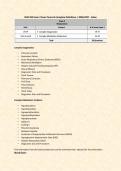
Complex Oxygenation
• Pulmonary Emboli
• Respiratory Failure
• Acute Respiratory Distress Syndrome (ARDS)
• Mechanical Ventilation
• Heparin Induced Thrombocytopenia (HIT)
• Pleural Effusion
• Diagnostics Tests and Procedures
• Chest Trauma
• Pulmonary Contusion
• Flail Chest
• Pneumothorax
• Hemothorax
• Chest Tubes
• Diagnostics Tests and Procedures
Complex Metabolism Problems
• Hypothyroidism
• Hyperthyroidism
• Hypoparathyroidism
• Hyperparathyroidism
• Hypophysectomy
• Addison’s
• Cushing’s
• Pheochromocytoma
• Diabetes Insipidus
• Syndrome of Inappropriate Antidiuretic Hormone (SIADH)
• Hyperglycemic-Hyperosmolar State (HHS)
• Diabetic Ketoacidosis (DKA)
• Diagnostics test and Procedures
Find information from the book provided and use the worksheet that I attached for that information
Blood Gases
,pH = 7.35 – 7.45
• pCO2 = 35 – 45
• The respiratory component.
• HCO3 = 22 – 28
• The metabolic component.
• Increased CO2 = acid build-up or acidosis.
• Increased HCO3 = alkaline build-up or alkalosis.
Key Points on ABG Interpretation:
Acidosis:
• Definition: Acidosis occurs when the arterial blood pH is less than 7.35.
• Types: o Metabolic Acidosis:
Low pH (<7.35)
Low bicarbonate (HCO₃ <22 mEq/L)⁻
Normal or slightly low PaCO₂ (35–40 mmHg)
Causes include diabetic ketoacidosis, renal failure, and prolonged
diarrhea. o Respiratory Acidosis:
Low pH (<7.35)
High PaCO₂ (>45 mmHg, sometimes exceeding 100 mmHg)
Normal or elevated bicarbonate in chronic cases due to renal
compensation.
Common in COPD, pneumonia, and respiratory depression.
Alkalosis:
• Definition: Alkalosis occurs when the arterial blood pH is greater than 7.45.
• Types: o Metabolic Alkalosis:
High pH (>7.45)
High bicarbonate (HCO₃ >28 mEq/L)⁻
Normal oxygen and carbon dioxide levels.
Causes include excessive antacid use, vomiting, and diuretics.
o Respiratory Alkalosis:
High pH (>7.45)
Low PaCO₂ (<35 mmHg)
Often caused by hyperventilation, anxiety, or high altitudes.
Nursing Interventions for Acid-Base Imbalances:
• Acidosis:
o Metabolic Acidosis: Provide rehydration, administer insulin (if due to
ketoacidosis), and use bicarbonate therapy if needed. o Respiratory
, Acidosis: Administer bronchodilators, provide oxygen therapy, and
monitor respiratory status.
• Alkalosis:
o Prevent further losses of electrolytes (H , K , Cl , Ca² )⁺ ⁺ ⁻ ⁺ . o Metabolic
Alkalosis: Correct underlying causes, provide fluids and electrolyte
replacement. o Respiratory Alkalosis: Reassure the patient, encourage
breathing into a paper bag, and adjust ventilator settings if necessary
Pulmonary Emboli (PE) - A collection of particulate matter (solids, liquids, or air) that
enters venous circulation and lodges in the pulmonary vessels, obstructing blood
flow and leading to hypoxia
Chapter & Page Number: Chapter 26, Page 597
Anticipated/Expected Findings:
• Sudden onset of dyspnea
• Pleuritic chest pain
• Tachypnea
• Tachycardia
• Hemoptysis
• Decreased oxygen saturation
• Anxiety and restlessness
• Petechiae
Complications/Unexpected Findings:
• Cardiac arrest
• Hypotension due to obstructive shock
• Right-sided heart failure (acute cor pulmonale)
• Severe hypoxemia
Past Medical History/Risk Factors:
• Recent surgery (especially orthopedic or abdominal)
• Prolonged immobility or bed rest
• Deep vein thrombosis (DVT) Pregnancy
, Oral contraceptive or hormone therapy use
• Cancer
• Smoking
Clinical Manifestations:
• VS: Increased heart rate (tachycardia), decreased SpO₂, hypotension in severe cases
• Cardiac: Chest pain, possible arrhythmias
• GI: Nausea, vomiting (less common)
• GU: No direct effects
• Integumentary: Pallor or cyanosis in severe cases
• Musculoskeletal: No direct effects
• Neurological: Anxiety, dizziness, syncope in severe cases
• Respiratory: Dyspnea, tachypnea, pleuritic chest pain, possible hemoptysis Labs:
• WBC: Normal or slightly elevated if inflammatory response is triggered
• Hgb/Hct: Normal unless there is an associated bleeding disorder
• Platelets: May be increased due to hypercoagulability
• aPTT/PT/INR: Elevated if anticoagulation therapy is initiated
• D-Dimer: Elevated (indicative of clot presence)
• ABGs: May show respiratory alkalosis initially (low CO₂), then hypoxemia (low PaO₂)
Cultures:
• Not applicable unless sepsis is suspected
Diagnostic/Imaging:
• CT Pulmonary Angiography: Gold standard for diagnosis
• V/Q Scan: Used if contrast allergy is present
• Doppler Ultrasound: To check for DVT
• Chest X-ray: To rule out other causes of symptoms
• Echocardiogram: To assess right heart strain
Provider Orders (Diet, IVF, Medications):
• Diet: Regular, unless contraindicated
• IV Fluids: Normal saline or lactated Ringer’s for hemodynamic support Medications:
o Heparin infusion or Low-Molecular-Weight Heparin (LMWH)
o Warfarin for long-term anticoagulation o Thrombolytic therapy (tPA) in
massive PE o Oxygen therapy as needed o Lovenox (enoxaparin)
o Arixtra (fondaparinux) o Xarelto (rivaroxaban)
Complex Oxygenation
• Pulmonary Emboli
• Respiratory Failure
• Acute Respiratory Distress Syndrome (ARDS)
• Mechanical Ventilation
• Heparin Induced Thrombocytopenia (HIT)
• Pleural Effusion
• Diagnostics Tests and Procedures
• Chest Trauma
• Pulmonary Contusion
• Flail Chest
• Pneumothorax
• Hemothorax
• Chest Tubes
• Diagnostics Tests and Procedures
Complex Metabolism Problems
• Hypothyroidism
• Hyperthyroidism
• Hypoparathyroidism
• Hyperparathyroidism
• Hypophysectomy
• Addison’s
• Cushing’s
• Pheochromocytoma
• Diabetes Insipidus
• Syndrome of Inappropriate Antidiuretic Hormone (SIADH)
• Hyperglycemic-Hyperosmolar State (HHS)
• Diabetic Ketoacidosis (DKA)
• Diagnostics test and Procedures
Find information from the book provided and use the worksheet that I attached for that information
Blood Gases
,pH = 7.35 – 7.45
• pCO2 = 35 – 45
• The respiratory component.
• HCO3 = 22 – 28
• The metabolic component.
• Increased CO2 = acid build-up or acidosis.
• Increased HCO3 = alkaline build-up or alkalosis.
Key Points on ABG Interpretation:
Acidosis:
• Definition: Acidosis occurs when the arterial blood pH is less than 7.35.
• Types: o Metabolic Acidosis:
Low pH (<7.35)
Low bicarbonate (HCO₃ <22 mEq/L)⁻
Normal or slightly low PaCO₂ (35–40 mmHg)
Causes include diabetic ketoacidosis, renal failure, and prolonged
diarrhea. o Respiratory Acidosis:
Low pH (<7.35)
High PaCO₂ (>45 mmHg, sometimes exceeding 100 mmHg)
Normal or elevated bicarbonate in chronic cases due to renal
compensation.
Common in COPD, pneumonia, and respiratory depression.
Alkalosis:
• Definition: Alkalosis occurs when the arterial blood pH is greater than 7.45.
• Types: o Metabolic Alkalosis:
High pH (>7.45)
High bicarbonate (HCO₃ >28 mEq/L)⁻
Normal oxygen and carbon dioxide levels.
Causes include excessive antacid use, vomiting, and diuretics.
o Respiratory Alkalosis:
High pH (>7.45)
Low PaCO₂ (<35 mmHg)
Often caused by hyperventilation, anxiety, or high altitudes.
Nursing Interventions for Acid-Base Imbalances:
• Acidosis:
o Metabolic Acidosis: Provide rehydration, administer insulin (if due to
ketoacidosis), and use bicarbonate therapy if needed. o Respiratory
, Acidosis: Administer bronchodilators, provide oxygen therapy, and
monitor respiratory status.
• Alkalosis:
o Prevent further losses of electrolytes (H , K , Cl , Ca² )⁺ ⁺ ⁻ ⁺ . o Metabolic
Alkalosis: Correct underlying causes, provide fluids and electrolyte
replacement. o Respiratory Alkalosis: Reassure the patient, encourage
breathing into a paper bag, and adjust ventilator settings if necessary
Pulmonary Emboli (PE) - A collection of particulate matter (solids, liquids, or air) that
enters venous circulation and lodges in the pulmonary vessels, obstructing blood
flow and leading to hypoxia
Chapter & Page Number: Chapter 26, Page 597
Anticipated/Expected Findings:
• Sudden onset of dyspnea
• Pleuritic chest pain
• Tachypnea
• Tachycardia
• Hemoptysis
• Decreased oxygen saturation
• Anxiety and restlessness
• Petechiae
Complications/Unexpected Findings:
• Cardiac arrest
• Hypotension due to obstructive shock
• Right-sided heart failure (acute cor pulmonale)
• Severe hypoxemia
Past Medical History/Risk Factors:
• Recent surgery (especially orthopedic or abdominal)
• Prolonged immobility or bed rest
• Deep vein thrombosis (DVT) Pregnancy
, Oral contraceptive or hormone therapy use
• Cancer
• Smoking
Clinical Manifestations:
• VS: Increased heart rate (tachycardia), decreased SpO₂, hypotension in severe cases
• Cardiac: Chest pain, possible arrhythmias
• GI: Nausea, vomiting (less common)
• GU: No direct effects
• Integumentary: Pallor or cyanosis in severe cases
• Musculoskeletal: No direct effects
• Neurological: Anxiety, dizziness, syncope in severe cases
• Respiratory: Dyspnea, tachypnea, pleuritic chest pain, possible hemoptysis Labs:
• WBC: Normal or slightly elevated if inflammatory response is triggered
• Hgb/Hct: Normal unless there is an associated bleeding disorder
• Platelets: May be increased due to hypercoagulability
• aPTT/PT/INR: Elevated if anticoagulation therapy is initiated
• D-Dimer: Elevated (indicative of clot presence)
• ABGs: May show respiratory alkalosis initially (low CO₂), then hypoxemia (low PaO₂)
Cultures:
• Not applicable unless sepsis is suspected
Diagnostic/Imaging:
• CT Pulmonary Angiography: Gold standard for diagnosis
• V/Q Scan: Used if contrast allergy is present
• Doppler Ultrasound: To check for DVT
• Chest X-ray: To rule out other causes of symptoms
• Echocardiogram: To assess right heart strain
Provider Orders (Diet, IVF, Medications):
• Diet: Regular, unless contraindicated
• IV Fluids: Normal saline or lactated Ringer’s for hemodynamic support Medications:
o Heparin infusion or Low-Molecular-Weight Heparin (LMWH)
o Warfarin for long-term anticoagulation o Thrombolytic therapy (tPA) in
massive PE o Oxygen therapy as needed o Lovenox (enoxaparin)
o Arixtra (fondaparinux) o Xarelto (rivaroxaban)