
TOES
PROJECTION PATIENT POSITION REFERENCE POINT CENTRAL RAY STRUCTURE ER ADDITIONAL INFO
SHOWN
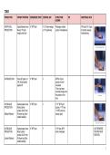
AP/AP AXIAL Supine/Seated; knee 3rd MTP joint ┴ (15o foam wedge) Phalanges & distal AP Axial (15o): Open
PROJECTION flexed; 15o foam or 15o posteriorly portion of metatarsals IP joints & reduces
wedge under foot foreshortening
PA PROJECTION Prone (IP joints // to 3rd MTP joint ┴ MTP & IP joint
CR); dorsal aspect spaces are well
against IR visualized
•The x-ray beam
coincides closely with
the position of the
toes
AP OBLIQUE Supine/seated; knee 3rd MTP joint ┴ 2nd-5th MTP joint
PROJECTION flexed; plantar spaces; 1st-3rd toes
surface 30-45o from •1st MTP joint (not
(Medial Rotation) IR (lower leg & foot always open)
rotated medially )
AP OBLIQUE Supine/seated; knee 3rd MTP joint ┴ 3rd-5th toes; MTP JUST REVERSE
PROJECTION flexed; plantar joints overlapped THE PREVIOUS
surface 30-45o from POSITION
(Lateral Rotation) IR (lower leg & foot
rotated laterally)
1
,PA OBLIQUE Lateral recumbent 3rd MTP joint ┴ 2nd-5th MTP joint
PROJECTION (affected side down); spaces; 1st-3rd toes
affected limb partially •1st MTP joint (not
(Medial Rotation) extended; ball of foot always open)
30o to horizontal
LATERAL Lateral recumbent IP joint (1st toe); ┴ Phalanges in profile;
PROJECTION (unaffected side proximal IP joint open IP joints spaces
down); toe in true (2nd-4th toes) •Lateromedial: 1st-2nd
(Mediolateral/Latero lateral; use 4x4 toes
medial Projection) gauze pad or tape (to •Mediolateral: 3rd-5th
separate the toes) toes
SESAMOIDS
PROJECTION PATIENT POSITION REFERENCE POINT CENTRAL RAY STRUCTURE ER ADDITIONAL INFO
SHOWN
TANGENTIAL Prone; dorsiflex great 1st MTP joint ┴ MT head &
PROJECTION toe; ankle elevated; sesamoids in profile
ball of foot ┴ to IR
LEWIS METHOD
2
,HOLLY METHOD Seated (more 1st MTP head ┴ MT head &
comfortable); plantar sesamoids in profile
surface 75o to IR; toe
flexed & hold w/ strip
gauze bandage;
medial border of foot
┴ to IR
CAUSTON METHOD Lateral recumbent Prominence of 1st 40o toward the heel Sesamoids projection
(unaffected side MTP joint axiolaterally with
down); knees flexed; slight overlap
limb partially
extended; foot in
lateral position; 1st
MTP joint ┴ to IR
FOOT
PROJECTION PATIENT POSITION REFERENCE POINT CENTRAL RAY STRUCTURE ER ADDITIONAL INFO
SHOWN
AP/AP AXIAL Supine; knee flexed; 3rd MTP base ┴ or 10o posteriorly MT & Tarsal (┴); TMT ● For localizing 10o Angulation:
PROJECTION plantar surface joint (10o) foreign bodies reduces
against IR ● Location of foreshortening of
fragments in fx metatarsals
of metatarsals &
anterior tarsals
● General surveys
of bones of the
foot
3
, AP OBLIQUE Supine; knee flexed; 3rd MTP base ┴ ● Cuboid in profile
PROJECTION leg rotated medially; ● Sinus tarsi (well
plantar surface of foot demonstrated)
(Medial Rotation) 30o to IR ● Interspaces b/n:
- cuboid &
calcaneus;
- cuboid & 4th &
5th MT
- Talus &
navicular
bone
AP OBLIQUE Supine; knee flexed; 3rd MTP base ┴ ● Navicular in
PROJECTION leg rotated laterally; profile
plantar surface of foot ● Interspaces b/n:
(Lateral Rotation) 30o to IR - 1st & 2nd MT
- Medial &
intermediate
cuneiforms
● Separates 1st-2nd
MT bases
LATERAL Dorsiflex foot (┴ to 3rd MT base ┴ Entire foot in profile ● For localizing more comfortable for
PROJECTION lower leg); leg & foot foreign body patient
in lateral position; ● Degree of
(Mediolateral lateral side of foot anterior &
Projection) against IR; posterior
displacement of
fx
4
PROJECTION PATIENT POSITION REFERENCE POINT CENTRAL RAY STRUCTURE ER ADDITIONAL INFO
SHOWN
AP/AP AXIAL Supine/Seated; knee 3rd MTP joint ┴ (15o foam wedge) Phalanges & distal AP Axial (15o): Open
PROJECTION flexed; 15o foam or 15o posteriorly portion of metatarsals IP joints & reduces
wedge under foot foreshortening
PA PROJECTION Prone (IP joints // to 3rd MTP joint ┴ MTP & IP joint
CR); dorsal aspect spaces are well
against IR visualized
•The x-ray beam
coincides closely with
the position of the
toes
AP OBLIQUE Supine/seated; knee 3rd MTP joint ┴ 2nd-5th MTP joint
PROJECTION flexed; plantar spaces; 1st-3rd toes
surface 30-45o from •1st MTP joint (not
(Medial Rotation) IR (lower leg & foot always open)
rotated medially )
AP OBLIQUE Supine/seated; knee 3rd MTP joint ┴ 3rd-5th toes; MTP JUST REVERSE
PROJECTION flexed; plantar joints overlapped THE PREVIOUS
surface 30-45o from POSITION
(Lateral Rotation) IR (lower leg & foot
rotated laterally)
1
,PA OBLIQUE Lateral recumbent 3rd MTP joint ┴ 2nd-5th MTP joint
PROJECTION (affected side down); spaces; 1st-3rd toes
affected limb partially •1st MTP joint (not
(Medial Rotation) extended; ball of foot always open)
30o to horizontal
LATERAL Lateral recumbent IP joint (1st toe); ┴ Phalanges in profile;
PROJECTION (unaffected side proximal IP joint open IP joints spaces
down); toe in true (2nd-4th toes) •Lateromedial: 1st-2nd
(Mediolateral/Latero lateral; use 4x4 toes
medial Projection) gauze pad or tape (to •Mediolateral: 3rd-5th
separate the toes) toes
SESAMOIDS
PROJECTION PATIENT POSITION REFERENCE POINT CENTRAL RAY STRUCTURE ER ADDITIONAL INFO
SHOWN
TANGENTIAL Prone; dorsiflex great 1st MTP joint ┴ MT head &
PROJECTION toe; ankle elevated; sesamoids in profile
ball of foot ┴ to IR
LEWIS METHOD
2
,HOLLY METHOD Seated (more 1st MTP head ┴ MT head &
comfortable); plantar sesamoids in profile
surface 75o to IR; toe
flexed & hold w/ strip
gauze bandage;
medial border of foot
┴ to IR
CAUSTON METHOD Lateral recumbent Prominence of 1st 40o toward the heel Sesamoids projection
(unaffected side MTP joint axiolaterally with
down); knees flexed; slight overlap
limb partially
extended; foot in
lateral position; 1st
MTP joint ┴ to IR
FOOT
PROJECTION PATIENT POSITION REFERENCE POINT CENTRAL RAY STRUCTURE ER ADDITIONAL INFO
SHOWN
AP/AP AXIAL Supine; knee flexed; 3rd MTP base ┴ or 10o posteriorly MT & Tarsal (┴); TMT ● For localizing 10o Angulation:
PROJECTION plantar surface joint (10o) foreign bodies reduces
against IR ● Location of foreshortening of
fragments in fx metatarsals
of metatarsals &
anterior tarsals
● General surveys
of bones of the
foot
3
, AP OBLIQUE Supine; knee flexed; 3rd MTP base ┴ ● Cuboid in profile
PROJECTION leg rotated medially; ● Sinus tarsi (well
plantar surface of foot demonstrated)
(Medial Rotation) 30o to IR ● Interspaces b/n:
- cuboid &
calcaneus;
- cuboid & 4th &
5th MT
- Talus &
navicular
bone
AP OBLIQUE Supine; knee flexed; 3rd MTP base ┴ ● Navicular in
PROJECTION leg rotated laterally; profile
plantar surface of foot ● Interspaces b/n:
(Lateral Rotation) 30o to IR - 1st & 2nd MT
- Medial &
intermediate
cuneiforms
● Separates 1st-2nd
MT bases
LATERAL Dorsiflex foot (┴ to 3rd MT base ┴ Entire foot in profile ● For localizing more comfortable for
PROJECTION lower leg); leg & foot foreign body patient
in lateral position; ● Degree of
(Mediolateral lateral side of foot anterior &
Projection) against IR; posterior
displacement of
fx
4


