
Diagnostiek en behandeling van evenwichtsstoornissen
1. Vestibulair systeem – balance & motion management
1.1. Doel en functie van het vestibulair systeem (evenwichtsorgaan)
• Functies van het evenwichtsorgaan:
o Ruimtelijke oriëntatie
o Beeld-/blikstabilisatie (voorbeeld met vinger- en hoofdbeweging)
o Houdingsstabilisatie
• Rechtopstaande houding bewaren
o Via vestibulo-spinale reflexen (VSR): reflexmatige houdingscorrecties ter compensatie van
hoofdbewegingen
o Vestibulospinaal systeem: vestibulum à vestibulaire kernen in de hersenstam à neuronen in ruggenmerg
à spieren
o Via vestibulo-collische reflex (VCR): hoofd/nek stabiel houden bij bewegingen
• Stabiel beeld op de omgeving bewaren
o Via vestibulo-oculaire reflexen (VOR): compensatoire oogbewegingen bij hoofdbewegingen
1.2. Onderzoeksmethoden
• Vestibulo-spinale reflexen:
o Evaluatie door het onderzoeken van corrigerende bewegingen van het lichaam
o Techniek: STABILOMETRIE of POSTUROGRAFIE
• Vestibulo-oculaire reflexen:
o Evaluatie door het onderzoeken van corrigerende oogbewegingen
o Techniek: VIDEONYSTAGMOGRAFIE (VNG) of ELEKTRONYSTAGMOGRAFIE (ENG)
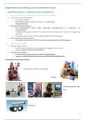
Voorbeelden van onderzoeksmethoden:
Posturografie: onderzoek + behandeling
®Equitest
Videonystagmografie (VNG)
®Difra
Frenzel bril
1
,1.3. Bouw van het vestibulair systeem
Detectie van hoek- of lineaire versnellingen (nooit zuiver lineair of hoek)
Anatomische bouw:
• Semicirculaire kanalen – voor draaibewegingen
• Sacculus & utriculus – voor rechtlijnige bewegingen en hoofdstand
à Het CZS verwerkt alle prikkels
Lokalisatie:
• Het ovale venster, dat door de voetplaat van de stapes wordt afgesloten, leidt naar het vestibulum of voorhof
• Aan de voorzijde (neus) van het vestibulum ligt de cochlea
• Aan de achterzijde (achterhoofd) monden de semicirculaire kanalen uit
Labyrintstructuur (net zoals in de cochlea):
• Beenderig labyrint: bevat perilymfe (heldere vloeistof)
• Membraneus labyrint: bevat endolymfe (slijmachtige vloeistof)
• Het membraneus labyrint bestaat uit een stelsel van kanalen en vliezige
blaasjes, omgeven door een hard beenderig kapsel
Semicirculaire kanalen:
• Drie kanalen:
o Eén voorste of anterieur SCK
o Eén achterste of posterieur SCK
o Eén horizontaal of lateraal SCK
• Functie: detectie van rotatoire hoofdbewegingen in het vlak van elk kanaal (relevant bij o.a. BPPV)
2
, • Aan de basis van elk semicirculair kanaal bevindt zich een verdikking: de ampulla
• In de ampulla ligt het zintuiglijk weefsel: de crista ampullaris
• De crista ampullaris met sensorische haarcellen ligt ingebed in de cupula (gelatineuze massa)
o Functie: detectie van stroming van de endolymfe
Innervatie
Utriculus en sacculus (vliezige zakjes):
• = otolietorganen (evenwichtssteentjes)
• In de wand van deze organen ligt het zintuiglijk weefsel: de macula
o De macula utriculi
o De macula sacculi
Structuur van de macula:
• Bestaat uit:
o Steuncellen
o Twee soorten zintuigcellen:
§ 70 à 80 stereocilia (cfr cochlea)
§ 1 kinocilium (langste haarcel)
• Boven op de zintuigcellen ligt het otolietmembraan:
o Geleiachtig membraan
o Bevat otolieten (calciumcarbonaatkristallen)
3
, 1.4. Balance & motion management
1.4.1. Het oog
1.4.2. Controle van oogbewegingen
Functies van de extra-oculaire oogspieren:
• Adductie: oog beweegt naar de neus (binnenzijde) – horizontale beweging (links-rechts)
• Abductie: oog beweegt naar de slaap (buitenzijde) – horizontale beweging (links-rechts)
• Elevatie: oog beweegt omhoog
• Depressie: oog beweegt omlaag
• Intorsie: oog draait naar binnen, de bovenpool van de oogbol roteert naar de neus – draaiende beweging
• Extorsie: oog draait naar buiten, bovenpool van de oogbol roteert naar de slaap – draaiende beweging
Oculomotore zenuwbanen:
• Nervus oculomotorius (III)
• Nervus trochlearis (IV) – m. obliquus superior
• Nervus abducens (VI) – m. rectus lateralis
à Ezelsbruggetje: LR6 SO4 rest III
• Laterale Rectus = VI (n. abducens)
• Superior Oblique = IV (n. trochlearis)
• De rest = III (n. oculomotorius)
4


