

C
WAVE -
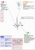
Nat QRS COMPLEX (Roms-3 small blocks) -
Nat
>
-
atrial depolarization >
-
ventricular depolarization
>
depolarization starts in the SA nade > narrow 9RS /IZOms
- -
spreads throughout the atria until it >
-
wide QRS) 120ms ,
reaches the AV nade So O-a 11s)
. .
>
-
in general men have a
, larger amplitude than women & young people
>
-
morphology : have higher amplitudes than the elderly
Sinus P waves should be up in 1 11 3 in limb leads leads is
>
voltage > Smm & < 10mm in precardial limb
- -
,
Vp- Vo 3 down in a VR s either the or-ve a bnormal
in the other leads >
-
small amounts of pericardial fluid are normal , with a larger effusion
-
look for evidence of atrial enlargement you will see dampening effects
>
-
if there are no p waves , are there fibrillation or
f luther waves ?
RL -
diffuse I paints are associated with early repolarization ,
LVH with strain
&
is pericarditis
-
AMIs sometimes have a diffuse J point especially
,
when there is tombstoning
Xipsegment
↓
1 7
↓
6
J point
M
ST
PR segment
Q
seg ment
S
T WAVE-13 +
>
-
ventricular repolarization
>
-
asymmetry is the normal presentation of the T wave
-
symmetrical T waves are found in ischemia , electrolyte
INTERVALS >
-
4) Inarmalities
peakedT wave :
& SNS problems
hyperkalaemia Stall is narrow)
>
-
broad T waves are found in CNS events (intercranial hemarrhage)
PR 120-200ms 7)20ms : abnormal connection between >
-
area that reflects ischemic insult ar injury to the myasardium
a tria 3 ventricles >
-
ischemia : ST depression 3 T waves in apposite direction from normal
> 200ms :
delayed conduction through >
-
myocardial injury ST elevation withoutT wave changes
:
,
Av mode > T waves
-
are usually the in leads 1 11 V3-Vo . ,
> T waves
-
are usually - ve in lead aVR
> Somm
height in limb leads
-
& RS < 120 ms > 120ms : abnormal sequence of ventricular >
-
> 12mm
height in precordial leads
depolarization ,
or excessive ventricular
mass
QT X RR interval >↳ RR interval :
delayed ventricular
repolarization
, ECG PAPER SRULES OF ECGS ATRIAL VENTRICULAR RELATIONSHIP
1 NEVER trust the computer is the PR interval
>
-
normal (120-200ms) ,
short or prolonged ?
-
exceptions : -
consistently >200ms indicative of 1st degree AU block
>
-
computer calculated HR is correct 99 9 % of the time . >
-
does the PR interval change ?
> if
-
the computer's overall impression is "normal ECG" ,
>
-
is there a 1 : / correspondence between P waves 3ORS complexes ?
then the ECG is highly likely to be normal >
-
do P waves come before/after the ORS complexes with which they are associated ?
2 ALWAYS examine the ECG systematically
-
rhythm -
> fast ,
slow or normal ? regular or irregular ?
>
-
narrow or wide CORS complex) ?
> what is the atrial activity ? IP waves)
-
>
-
what is the relationship between atrials ventricular
activity ? IPR interval relationship of P wave BORS)
-
QRS axis ORS morphology
-
ST Segment 3 T wave morphology
-- > ST
-
segment depressions or elevations
T wave flattening or inversion
- "Big box" Smm >
-
:
=
200ms >
-
examine OT interval
7) 1 "small box" = 1 mm =
40ms 3 consider the clinical context when looking at ECG impression
: S big boxes = 1S 4 compare current previous ECGs (if available)
>
-
evaluating ischemia and/or infarction
5 incorporate ECG impression into the generation of the
differential diagnosis
DETERMINING RATE DETERMINING AXIS
>
-
normal resting heart rate : So-90bpm
> max
-
Sinus rate = 220-patient age
.
>
-
rate of 140-150 bpm suggests possibility of atrial THE QRS AXIS
f lutter with 2 : 1 AV block
represents the average direction of electrical
-
activity in the heart during ventricular depolarization
THE 300 RULE -
deviation can provide insight into chamber
enlargement abnormalities of the conduction system
beats-[s] [sbigboxes] [es]
, ,
. .
myocardial infarctions ,
3 the origin of some
arrhythmias
HR (bpm) =
300
NON-PATHOLOGIC FACTORS AFFECTING QRS AXIS
# big boxes between beats
-
age : moves leftward as an individual ages
* cannot apply this rule when the ventricular rhythm is -
body type : tall s thin -> verticle axis
irregular
short Obese >-
leftward axis
THE 10 SECOND RULE
HR (bpm) = # of beats across ECG X 6
(gives appearance of widened a
>look fordelta
-
waves a
WAVE -
Nat QRS COMPLEX (Roms-3 small blocks) -
Nat
>
-
atrial depolarization >
-
ventricular depolarization
>
depolarization starts in the SA nade > narrow 9RS /IZOms
- -
spreads throughout the atria until it >
-
wide QRS) 120ms ,
reaches the AV nade So O-a 11s)
. .
>
-
in general men have a
, larger amplitude than women & young people
>
-
morphology : have higher amplitudes than the elderly
Sinus P waves should be up in 1 11 3 in limb leads leads is
>
voltage > Smm & < 10mm in precardial limb
- -
,
Vp- Vo 3 down in a VR s either the or-ve a bnormal
in the other leads >
-
small amounts of pericardial fluid are normal , with a larger effusion
-
look for evidence of atrial enlargement you will see dampening effects
>
-
if there are no p waves , are there fibrillation or
f luther waves ?
RL -
diffuse I paints are associated with early repolarization ,
LVH with strain
&
is pericarditis
-
AMIs sometimes have a diffuse J point especially
,
when there is tombstoning
Xipsegment
↓
1 7
↓
6
J point
M
ST
PR segment
Q
seg ment
S
T WAVE-13 +
>
-
ventricular repolarization
>
-
asymmetry is the normal presentation of the T wave
-
symmetrical T waves are found in ischemia , electrolyte
INTERVALS >
-
4) Inarmalities
peakedT wave :
& SNS problems
hyperkalaemia Stall is narrow)
>
-
broad T waves are found in CNS events (intercranial hemarrhage)
PR 120-200ms 7)20ms : abnormal connection between >
-
area that reflects ischemic insult ar injury to the myasardium
a tria 3 ventricles >
-
ischemia : ST depression 3 T waves in apposite direction from normal
> 200ms :
delayed conduction through >
-
myocardial injury ST elevation withoutT wave changes
:
,
Av mode > T waves
-
are usually the in leads 1 11 V3-Vo . ,
> T waves
-
are usually - ve in lead aVR
> Somm
height in limb leads
-
& RS < 120 ms > 120ms : abnormal sequence of ventricular >
-
> 12mm
height in precordial leads
depolarization ,
or excessive ventricular
mass
QT X RR interval >↳ RR interval :
delayed ventricular
repolarization
, ECG PAPER SRULES OF ECGS ATRIAL VENTRICULAR RELATIONSHIP
1 NEVER trust the computer is the PR interval
>
-
normal (120-200ms) ,
short or prolonged ?
-
exceptions : -
consistently >200ms indicative of 1st degree AU block
>
-
computer calculated HR is correct 99 9 % of the time . >
-
does the PR interval change ?
> if
-
the computer's overall impression is "normal ECG" ,
>
-
is there a 1 : / correspondence between P waves 3ORS complexes ?
then the ECG is highly likely to be normal >
-
do P waves come before/after the ORS complexes with which they are associated ?
2 ALWAYS examine the ECG systematically
-
rhythm -
> fast ,
slow or normal ? regular or irregular ?
>
-
narrow or wide CORS complex) ?
> what is the atrial activity ? IP waves)
-
>
-
what is the relationship between atrials ventricular
activity ? IPR interval relationship of P wave BORS)
-
QRS axis ORS morphology
-
ST Segment 3 T wave morphology
-- > ST
-
segment depressions or elevations
T wave flattening or inversion
- "Big box" Smm >
-
:
=
200ms >
-
examine OT interval
7) 1 "small box" = 1 mm =
40ms 3 consider the clinical context when looking at ECG impression
: S big boxes = 1S 4 compare current previous ECGs (if available)
>
-
evaluating ischemia and/or infarction
5 incorporate ECG impression into the generation of the
differential diagnosis
DETERMINING RATE DETERMINING AXIS
>
-
normal resting heart rate : So-90bpm
> max
-
Sinus rate = 220-patient age
.
>
-
rate of 140-150 bpm suggests possibility of atrial THE QRS AXIS
f lutter with 2 : 1 AV block
represents the average direction of electrical
-
activity in the heart during ventricular depolarization
THE 300 RULE -
deviation can provide insight into chamber
enlargement abnormalities of the conduction system
beats-[s] [sbigboxes] [es]
, ,
. .
myocardial infarctions ,
3 the origin of some
arrhythmias
HR (bpm) =
300
NON-PATHOLOGIC FACTORS AFFECTING QRS AXIS
# big boxes between beats
-
age : moves leftward as an individual ages
* cannot apply this rule when the ventricular rhythm is -
body type : tall s thin -> verticle axis
irregular
short Obese >-
leftward axis
THE 10 SECOND RULE
HR (bpm) = # of beats across ECG X 6
(gives appearance of widened a
>look fordelta
-
waves a


