
Topic 7: Run for Your Life | Pearson Edexcel Biology A (Salters-Nuffield) / 9BN0
Joints and movement
7.1 / muscles, tendons and bones work together; antagonistic pairs
Bones are moved at joints by skeletal muscles. A muscle is attached to bone by tendons. When a muscle
contracts it shortens and pulls on the bone; a muscle cannot push, so it can only be stretched back to its
original length when the opposing muscle contracts.
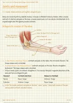
Antagonistic muscles at the elbow
humerus
biceps: the flexor (front of the upper arm)
triceps: the extensor
elbow: a hinge joint
(back of the upper arm)
tendon (to the radius)
radius
tendon (to the ulna)
ulna
To flex the arm, the biceps (flexor) contracts and pulls on the radius: the arm bends (flexion). The
triceps relaxes and is stretched.
To extend the arm, the triceps (extensor) contracts and pulls on the ulna: the arm straightens
(extension). The biceps relaxes and is stretched.
A flexor bends a joint; an extensor straightens it. Two muscles that pull in opposite directions at the
same joint are an antagonistic pair.
Movement Contracts (shortens) Relaxes (is stretched)
Flexion Flexor: biceps Extensor: triceps
Extension Extensor: triceps Flexor: biceps
Write "the muscle contracts and pulls on the bone", never "pushes". Name both muscles of the pair
and say which contracts and which relaxes.
Topic 7: Run for Your Life
,Topic 7: Run for Your Life | Pearson Edexcel Biology A (Salters-Nuffield) / 9BN0
Inside a synovial joint
7.1 / ligaments, cartilage and synovial fluid allow smooth movement
Joints such as the elbow, knee and hip are synovial joints: the bones move freely against each other. The
parts of the joint hold the bones in place while reducing friction between them.
A synovial joint in section
bone
joint capsule with ligaments
synovial membrane
cartilage
synovial fluid in the joint cavity
bone
Structure What it is Function
Tendon Strong, inelastic (non-elastic) cord of Joins muscle to bone; passes on the pull
collagen. of the muscle.
Ligament Strong, flexible and slightly elastic Joins bone to bone; holds the joint
band. together and controls its movement.
Cartilage Smooth tissue covering the ends of Acts as a shock absorber and stops the
the bones. bones rubbing together.
Synovial membrane Lining of the joint capsule. Secretes synovial fluid.
Synovial fluid Liquid filling the joint cavity. Lubricates the joint: reduces friction.
Tendon = muscle to bone and must not stretch, or the pull would be lost. Ligament = bone to bone and
must stretch a little to let the joint move.
Topic 7: Run for Your Life
,Topic 7: Run for Your Life | Pearson Edexcel Biology A (Salters-Nuffield) / 9BN0
Inside a muscle fibre: the sarcomere
7.2 / actin and myosin filaments give the banding pattern
A muscle fibre is packed with myofibrils. Each myofibril is a chain of repeating units called sarcomeres,
built from two protein filaments: thin actin and thick myosin. Their overlap gives the striped appearance.
One sarcomere: relaxed and contracted
Z line M line
Relaxed
actin (thin) sarcomere
myosin (thick) A band (length of myosin)
I band H zone
Contracted
sarcomere: shorter
I band: shorter A band: unchanged H zone: shorter
Region What it is When the muscle contracts
Z line End of the sarcomere; actin is attached to it. Z lines move closer together.
I band Light band: actin only. Gets shorter.
A band Dark band: the full length of the myosin. Stays the same length.
H zone Centre of the A band: myosin only. Gets shorter.
The filaments themselves do not shorten. Actin slides between the myosin, increasing the overlap, so
the sarcomere shortens.
Topic 7: Run for Your Life
Joints and movement
7.1 / muscles, tendons and bones work together; antagonistic pairs
Bones are moved at joints by skeletal muscles. A muscle is attached to bone by tendons. When a muscle
contracts it shortens and pulls on the bone; a muscle cannot push, so it can only be stretched back to its
original length when the opposing muscle contracts.
Antagonistic muscles at the elbow
humerus
biceps: the flexor (front of the upper arm)
triceps: the extensor
elbow: a hinge joint
(back of the upper arm)
tendon (to the radius)
radius
tendon (to the ulna)
ulna
To flex the arm, the biceps (flexor) contracts and pulls on the radius: the arm bends (flexion). The
triceps relaxes and is stretched.
To extend the arm, the triceps (extensor) contracts and pulls on the ulna: the arm straightens
(extension). The biceps relaxes and is stretched.
A flexor bends a joint; an extensor straightens it. Two muscles that pull in opposite directions at the
same joint are an antagonistic pair.
Movement Contracts (shortens) Relaxes (is stretched)
Flexion Flexor: biceps Extensor: triceps
Extension Extensor: triceps Flexor: biceps
Write "the muscle contracts and pulls on the bone", never "pushes". Name both muscles of the pair
and say which contracts and which relaxes.
Topic 7: Run for Your Life
,Topic 7: Run for Your Life | Pearson Edexcel Biology A (Salters-Nuffield) / 9BN0
Inside a synovial joint
7.1 / ligaments, cartilage and synovial fluid allow smooth movement
Joints such as the elbow, knee and hip are synovial joints: the bones move freely against each other. The
parts of the joint hold the bones in place while reducing friction between them.
A synovial joint in section
bone
joint capsule with ligaments
synovial membrane
cartilage
synovial fluid in the joint cavity
bone
Structure What it is Function
Tendon Strong, inelastic (non-elastic) cord of Joins muscle to bone; passes on the pull
collagen. of the muscle.
Ligament Strong, flexible and slightly elastic Joins bone to bone; holds the joint
band. together and controls its movement.
Cartilage Smooth tissue covering the ends of Acts as a shock absorber and stops the
the bones. bones rubbing together.
Synovial membrane Lining of the joint capsule. Secretes synovial fluid.
Synovial fluid Liquid filling the joint cavity. Lubricates the joint: reduces friction.
Tendon = muscle to bone and must not stretch, or the pull would be lost. Ligament = bone to bone and
must stretch a little to let the joint move.
Topic 7: Run for Your Life
,Topic 7: Run for Your Life | Pearson Edexcel Biology A (Salters-Nuffield) / 9BN0
Inside a muscle fibre: the sarcomere
7.2 / actin and myosin filaments give the banding pattern
A muscle fibre is packed with myofibrils. Each myofibril is a chain of repeating units called sarcomeres,
built from two protein filaments: thin actin and thick myosin. Their overlap gives the striped appearance.
One sarcomere: relaxed and contracted
Z line M line
Relaxed
actin (thin) sarcomere
myosin (thick) A band (length of myosin)
I band H zone
Contracted
sarcomere: shorter
I band: shorter A band: unchanged H zone: shorter
Region What it is When the muscle contracts
Z line End of the sarcomere; actin is attached to it. Z lines move closer together.
I band Light band: actin only. Gets shorter.
A band Dark band: the full length of the myosin. Stays the same length.
H zone Centre of the A band: myosin only. Gets shorter.
The filaments themselves do not shorten. Actin slides between the myosin, increasing the overlap, so
the sarcomere shortens.
Topic 7: Run for Your Life


