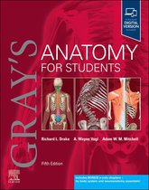
GRAY'S ANATOMY FOR STUDENTS
5TH EDITION
• AUTHOR(S)RICHARD L. DRAKE
TEST BANK
1)
Reference: Ch. 1 — The Body — What is anatomy?
Stem: A junior clinician is assessing a patient after a blunt
abdominal trauma. The clinician must decide whether to
palpate broadly across the abdomen or selectively examine
specific surface landmarks to avoid worsening a suspected
organ injury. Which anatomical reasoning best guides a targeted
physical exam to reduce risk and localize a possible intra-
abdominal injury?
Options:
A. Use generalized abdominal percussion only because surface
anatomy is too variable to localize organs.
B. Rely on mapped surface landmarks (e.g., McBurney’s point)
and regional anatomy to guide focused palpation.
C. Avoid surface landmarks and use only imaging because
,palpation always risks exacerbating internal injuries.
D. Palpate along dermatomal lines exclusively because organ
position correlates directly with single spinal cord segments.
Correct answer: B
Correct Answer Rationale (3–4 sentences):
Surface landmarks and regional anatomy provide clinically
useful guidance to underlying viscera and reduce unnecessary
manipulation of uninvolved areas. Gray’s emphasizes that
understanding topographical relationships (e.g., appendix near
McBurney’s point) enables focused, safer assessment and
better correlation with likely injured structures. Using regional
anatomy supports decision-making for urgent imaging or
limited-focused palpation.
Incorrect Answer Rationales (1–3 sentences each):
A. Overstates variability; while variation exists, standard surface
landmarks remain reliable for initial localization.
C. Imaging complements but does not fully replace targeted
physical exam—palpation can provide rapid bedside
information.
D. Dermatomes relate primarily to cutaneous sensation, not
precise organ boundaries, so they alone are insufficient for
organ localization.
Teaching Point: Surface anatomy + regional maps guide
focused, safer clinical exams.
,Citation: Drake, R. L. (2024). Gray’s Anatomy for Students (5th
Ed.). Ch. 1.
2)
Reference: Ch. 1 — The Body — Anatomical position &
directional terms
Stem: During a CT review, an intern describes a lesion as
“posterolateral to the proximal femur.” A surgeon asks for
clarification using standard anatomical terms. Which
description best translates the intern’s location into precise
anatomical orientation to guide incision planning?
Options:
A. The lesion lies behind and toward the midline of the proximal
femur.
B. The lesion lies behind and toward the outer side (away from
midline) of the proximal femur.
C. The lesion lies in front and medially near the distal femur.
D. The lesion lies superficially on the anterior surface of the
proximal femur.
Correct answer: B
Correct Answer Rationale (3–4 sentences):
“Posterolateral” combines posterior (behind) and lateral (away
from midline) — so the lesion is located posteriorly and laterally
relative to the proximal femur. Accurate use of directional terms
from the anatomical position prevents surgical misorientation
, and assists safe incision placement. Gray’s defines these terms
to standardize communication across clinicians and imaging.
Incorrect Answer Rationales (1–3 sentences each):
A. “Toward the midline” contradicts lateral; posterolateral is
away from midline.
C. Describes anterior and medial relationships and confuses
both direction and bone segment.
D. “Superficial anterior” is the opposite of posterior and does
not address lateral/medial orientation.
Teaching Point: Posterior = behind; lateral = away from midline
— combine terms precisely.
Citation: Drake, R. L. (2024). Gray’s Anatomy for Students (5th
Ed.). Ch. 1.
3)
Reference: Ch. 1 — The Body — Planes of section & imaging
orientation
Stem: A radiologist requests an MRI slice in the coronal plane to
evaluate a suspected rotator cuff tear. The trainee confuses
coronal with sagittal orientation. Which explanation best
distinguishes coronal from sagittal planes for selecting imaging
slices relevant to joint anatomy?
Options:
A. The coronal plane divides the body into left and right; it’s
5TH EDITION
• AUTHOR(S)RICHARD L. DRAKE
TEST BANK
1)
Reference: Ch. 1 — The Body — What is anatomy?
Stem: A junior clinician is assessing a patient after a blunt
abdominal trauma. The clinician must decide whether to
palpate broadly across the abdomen or selectively examine
specific surface landmarks to avoid worsening a suspected
organ injury. Which anatomical reasoning best guides a targeted
physical exam to reduce risk and localize a possible intra-
abdominal injury?
Options:
A. Use generalized abdominal percussion only because surface
anatomy is too variable to localize organs.
B. Rely on mapped surface landmarks (e.g., McBurney’s point)
and regional anatomy to guide focused palpation.
C. Avoid surface landmarks and use only imaging because
,palpation always risks exacerbating internal injuries.
D. Palpate along dermatomal lines exclusively because organ
position correlates directly with single spinal cord segments.
Correct answer: B
Correct Answer Rationale (3–4 sentences):
Surface landmarks and regional anatomy provide clinically
useful guidance to underlying viscera and reduce unnecessary
manipulation of uninvolved areas. Gray’s emphasizes that
understanding topographical relationships (e.g., appendix near
McBurney’s point) enables focused, safer assessment and
better correlation with likely injured structures. Using regional
anatomy supports decision-making for urgent imaging or
limited-focused palpation.
Incorrect Answer Rationales (1–3 sentences each):
A. Overstates variability; while variation exists, standard surface
landmarks remain reliable for initial localization.
C. Imaging complements but does not fully replace targeted
physical exam—palpation can provide rapid bedside
information.
D. Dermatomes relate primarily to cutaneous sensation, not
precise organ boundaries, so they alone are insufficient for
organ localization.
Teaching Point: Surface anatomy + regional maps guide
focused, safer clinical exams.
,Citation: Drake, R. L. (2024). Gray’s Anatomy for Students (5th
Ed.). Ch. 1.
2)
Reference: Ch. 1 — The Body — Anatomical position &
directional terms
Stem: During a CT review, an intern describes a lesion as
“posterolateral to the proximal femur.” A surgeon asks for
clarification using standard anatomical terms. Which
description best translates the intern’s location into precise
anatomical orientation to guide incision planning?
Options:
A. The lesion lies behind and toward the midline of the proximal
femur.
B. The lesion lies behind and toward the outer side (away from
midline) of the proximal femur.
C. The lesion lies in front and medially near the distal femur.
D. The lesion lies superficially on the anterior surface of the
proximal femur.
Correct answer: B
Correct Answer Rationale (3–4 sentences):
“Posterolateral” combines posterior (behind) and lateral (away
from midline) — so the lesion is located posteriorly and laterally
relative to the proximal femur. Accurate use of directional terms
from the anatomical position prevents surgical misorientation
, and assists safe incision placement. Gray’s defines these terms
to standardize communication across clinicians and imaging.
Incorrect Answer Rationales (1–3 sentences each):
A. “Toward the midline” contradicts lateral; posterolateral is
away from midline.
C. Describes anterior and medial relationships and confuses
both direction and bone segment.
D. “Superficial anterior” is the opposite of posterior and does
not address lateral/medial orientation.
Teaching Point: Posterior = behind; lateral = away from midline
— combine terms precisely.
Citation: Drake, R. L. (2024). Gray’s Anatomy for Students (5th
Ed.). Ch. 1.
3)
Reference: Ch. 1 — The Body — Planes of section & imaging
orientation
Stem: A radiologist requests an MRI slice in the coronal plane to
evaluate a suspected rotator cuff tear. The trainee confuses
coronal with sagittal orientation. Which explanation best
distinguishes coronal from sagittal planes for selecting imaging
slices relevant to joint anatomy?
Options:
A. The coronal plane divides the body into left and right; it’s