w Adult Spine 201
Self-Assessment Examination 201
2015
AAOS
Yowr Sorefor Lifelong Orthopaedic learig
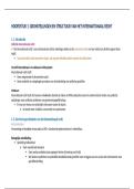
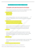
,1 - Figures 1 and 2 are CT scans obtained from a 68-year-old man who has had progressive neck pain and stiffness,
worsening gait imbalance, upper extremity weakness, early muscle fatigue, difficulty with fine motor control, and
difficulty with activities of daily living over the past few years. On physical examination, he has a wide based stiff legged
gait, generalized upper extremity weakness, dense sensory loss in the upper and lower extremities, and markedly brisk
reflexes. What is the most appropriate treatment for this patient?
Figure 1 Figure 2
A. Observation
B. Cervical epidural injections
C. Multilevel anterior cervical decompression and fusion
D. Posterior cervical laminoplasties from C3-6
Correct answer: D
This patient has progressive myelopathy secondary to ossification of the posterior longitudinal ligament. Diagnostic
imaging reveals multilevel cervical cord compression from C4-6. The patient has maintained reasonable cervical lordosis.
A posterior procedure such as multilevel laminoplasty decompresses the spine, is motion preserving, and has a low
complication rate. Observation and cervical epidural injections are not viable options in patients with progressive
myelopathy. Anterior cervical decompression, including corpectomy, is an option; however, anterior procedures have an
increased risk of complications such as dural tear or cerebrospinal fluid leak. The axial CT image shows a "double layer"
sign, which is consistent with dural ossification and increases the risk of dural injury with anterior decompression.
2 - When compared with posterior decompression and fusion, the addition of an interbody fusion for the treatment of
degenerative spondylolisthesis and stenosis has been shown to
A. result in increased patient functional outcome scores.
B. reduce the incidence of symptomatic pseudarthrosis.
, C. increase the length of hospital stay.
D. increase hospital costs.
Correct answer: D
The use of an interbody graft has been shown to increase hospital costs. Gottschalk and associates found no
change in Oswestry Disability Index (ODI) or 36-Item Short-Form Health Survey (SF-36) scores when
comparing patients fused using either posterior fusion or transforaminal interbody fusion. They also found no
change in fusion rates at 3 years after surgery. Carreon and associates showed some that using a posterior place
interbody transforaminal lumbar interbody fusion (TLIF) or posterior lumbar interbody fusion (PLIF) did result
in improved ODI and SF-6D scores but did not result in any change in EuroQol five dimensions questionnaire
(EQ-5D) scores. Using the EQ-5D data, they estimated that the use of an interbody graft becomes cost
prohibitive if the charges exceed $1,570 above the cost of a posterior fusion. The use of an interbody cage has
not been shown to increase hospital stay.
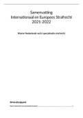
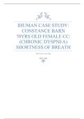
3 - Figures 1 and 2 are MRI images obtained from a 22-year-old man who fell from a 2-story building. On examination, he
has diminished rectal tone and urinary retention. If surgical stabilization is elected, what is the most biomechanically
stable option?
Figure 1 Figure 2
A. Sacral plating
B. Iliosacral screws
C. Iliosacral screws and lumbopelvic fixation
D. External fixation
, Correct answer: C
The fpatient fhas fa fU-shaped fsacral ffracture for fspondylopelvic fdissociation. fTreatment foptions ffor fthese ffractures
frange ffrom fpercutaneous fplacement fof filiosacral fscrews fto flumbopelvic ffixation f(lumbar fpedicle fscrews fand filiac
fscrews).
Lumbopelvic ffixation fcan fbe fsupplemented fby filiosacral fscrews, fwhich fhas fbeen ftermed ftriangular fosteosynthesis.
fBiomechanical fstudies fhave fshown fthat filiosacral fscrews fwith flumbopelvic ffixation—or ftriangular fosteosynthesis—is
fthe fmost fstable fconstruct fwhen fcompared fwith filiosacral fscrews falone. fThe fadvantage fof flumbopelvic ffixation fis
fthat fconcurrent fsacral flaminectomy fcan fbe fperformed, fwhich fis frecommend fin fthis fpatient fbecause fof fhis
fneurologic fsymptoms. fExternal ffixation for fsacral fplating fplay fminimal froles fin fU-shaped fsacral ffractures.
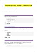
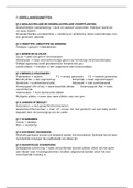
4 - fClinical fSituation
Figure f1 fshows fa fCT ffrom fthe fcervical fspine fof fan f85-year-old fwoman fwho ffell ffrom fa fstanding fheight f1
fweek fearlier. fShe fis findependent fand fambulatory fand fresides fin fan fassisted fliving ffacility. fShe freports
fpersistent fneck fpain fbut fdenies farm fpain for fweakness. fShe fis fneurologically fintact.
Fractures fin fthis fregion fof fC2 fhave fa fhigh frisk fof
Figure 1
A. spinal fcord finjury.
B. union.
C. nonunion.
D. stroke.
Self-Assessment Examination 201
2015
AAOS
Yowr Sorefor Lifelong Orthopaedic learig
,1 - Figures 1 and 2 are CT scans obtained from a 68-year-old man who has had progressive neck pain and stiffness,
worsening gait imbalance, upper extremity weakness, early muscle fatigue, difficulty with fine motor control, and
difficulty with activities of daily living over the past few years. On physical examination, he has a wide based stiff legged
gait, generalized upper extremity weakness, dense sensory loss in the upper and lower extremities, and markedly brisk
reflexes. What is the most appropriate treatment for this patient?
Figure 1 Figure 2
A. Observation
B. Cervical epidural injections
C. Multilevel anterior cervical decompression and fusion
D. Posterior cervical laminoplasties from C3-6
Correct answer: D
This patient has progressive myelopathy secondary to ossification of the posterior longitudinal ligament. Diagnostic
imaging reveals multilevel cervical cord compression from C4-6. The patient has maintained reasonable cervical lordosis.
A posterior procedure such as multilevel laminoplasty decompresses the spine, is motion preserving, and has a low
complication rate. Observation and cervical epidural injections are not viable options in patients with progressive
myelopathy. Anterior cervical decompression, including corpectomy, is an option; however, anterior procedures have an
increased risk of complications such as dural tear or cerebrospinal fluid leak. The axial CT image shows a "double layer"
sign, which is consistent with dural ossification and increases the risk of dural injury with anterior decompression.
2 - When compared with posterior decompression and fusion, the addition of an interbody fusion for the treatment of
degenerative spondylolisthesis and stenosis has been shown to
A. result in increased patient functional outcome scores.
B. reduce the incidence of symptomatic pseudarthrosis.
, C. increase the length of hospital stay.
D. increase hospital costs.
Correct answer: D
The use of an interbody graft has been shown to increase hospital costs. Gottschalk and associates found no
change in Oswestry Disability Index (ODI) or 36-Item Short-Form Health Survey (SF-36) scores when
comparing patients fused using either posterior fusion or transforaminal interbody fusion. They also found no
change in fusion rates at 3 years after surgery. Carreon and associates showed some that using a posterior place
interbody transforaminal lumbar interbody fusion (TLIF) or posterior lumbar interbody fusion (PLIF) did result
in improved ODI and SF-6D scores but did not result in any change in EuroQol five dimensions questionnaire
(EQ-5D) scores. Using the EQ-5D data, they estimated that the use of an interbody graft becomes cost
prohibitive if the charges exceed $1,570 above the cost of a posterior fusion. The use of an interbody cage has
not been shown to increase hospital stay.
3 - Figures 1 and 2 are MRI images obtained from a 22-year-old man who fell from a 2-story building. On examination, he
has diminished rectal tone and urinary retention. If surgical stabilization is elected, what is the most biomechanically
stable option?
Figure 1 Figure 2
A. Sacral plating
B. Iliosacral screws
C. Iliosacral screws and lumbopelvic fixation
D. External fixation
, Correct answer: C
The fpatient fhas fa fU-shaped fsacral ffracture for fspondylopelvic fdissociation. fTreatment foptions ffor fthese ffractures
frange ffrom fpercutaneous fplacement fof filiosacral fscrews fto flumbopelvic ffixation f(lumbar fpedicle fscrews fand filiac
fscrews).
Lumbopelvic ffixation fcan fbe fsupplemented fby filiosacral fscrews, fwhich fhas fbeen ftermed ftriangular fosteosynthesis.
fBiomechanical fstudies fhave fshown fthat filiosacral fscrews fwith flumbopelvic ffixation—or ftriangular fosteosynthesis—is
fthe fmost fstable fconstruct fwhen fcompared fwith filiosacral fscrews falone. fThe fadvantage fof flumbopelvic ffixation fis
fthat fconcurrent fsacral flaminectomy fcan fbe fperformed, fwhich fis frecommend fin fthis fpatient fbecause fof fhis
fneurologic fsymptoms. fExternal ffixation for fsacral fplating fplay fminimal froles fin fU-shaped fsacral ffractures.
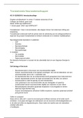
4 - fClinical fSituation
Figure f1 fshows fa fCT ffrom fthe fcervical fspine fof fan f85-year-old fwoman fwho ffell ffrom fa fstanding fheight f1
fweek fearlier. fShe fis findependent fand fambulatory fand fresides fin fan fassisted fliving ffacility. fShe freports
fpersistent fneck fpain fbut fdenies farm fpain for fweakness. fShe fis fneurologically fintact.
Fractures fin fthis fregion fof fC2 fhave fa fhigh frisk fof
Figure 1
A. spinal fcord finjury.
B. union.
C. nonunion.
D. stroke.