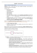
FINAL EXAM: Edapt Notes
Weeks 1-3
Hypersensitivity & Anemia
Key Concepts
, lOMoARcPSD|51648332
NR507 Edapts
Week 1
EDapt Module Questions
Hives (Urticaria) - Type 1 Allergic Reaction – dermal/skin manifestation.
Allergic Contact Dermatitis – Type 4 Allergic Reaction - an example of a Type
IV hypersensitivity reaction mediated by T-cells. When the individual comes in
contact with the antigen (e.g. poison ivy), an antigen complex is formed. On
subsequent exposure to the antigen, sensitized T-cells activate the inflammatory
process that causes the allergic contact dermatitis to appear.
Type 2 (Cytotoxic/tissue-specific) hypersensitivity reactions are mediated by: IgG
or IgM.
Which of the following are considered the “first responders” of the innate immune
system? Neutrophils appear first in any immune response.
Anaphylaxis is a Type 1 Allergic Reaction - Type 1 hypersensitivity reactions are
mediated by IgE and mast cells. An individual who is highly sensitized to the
antigen may experience anaphylaxis.
Damage occurs with ABO incompatibility because: Complement damages RBC
membrane causing cell lysis. Damage from ABO incompatibility occurs because
of the effects of complement on the RBC membrane that results in RBC lysis.
The diagnosis for an individual who presents to the office with sudden swollen
lips and eyes, shortness of breath and throat tightness after a bee sting is:
anaphylaxis. The symptoms are consistent with the life-threating condition,
anaphylaxis after being exposed. to a bee sting.
Which of the following assessment findings would be expected in a patient who
presents with urticaria? Eosinophilia. Eosinophils are present in the allergic
reaction.
Type IV cytotoxic hypersensitivity reactions are mediated by: T-cells.
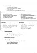
Types of Hypersensitivity Reactions
Downloaded by Benjamin Luca ()
, lOMoARcPSD|51648332
Type Mechanism Example Pathology
Mast cell degranulation
IgE action on mast
I. Hay fever results in an
cells
inflammatory response
Tissue-specific
destruction or
impairment
because of:
Antibody binding
followed by lysis
via complement
1-Complement
Antibody binding
damages RBC
followed by
1-ABO membrane and cells
macrophage
incompatibility lyse
II. phagocytosis
5-Graves' 5-Autoantibodies
Antibody binding
disease specific for thyroid
followed by
tissue impair receptor
neutrophil
for TSH
destruction
Antibody-
dependent cell
(NK)-mediated
cytotoxicity, or
Antireceptor
antibodies
III. Antigen-Antibody Raynaud’s Complex deposited in
complex deposited phenomenon small peripheral
in tissues vessels in cool
temperatures leading
to vasoconstriction and
Downloaded by Benjamin Luca ()
, lOMoARcPSD|51648332
blocked circulation
Contact
Cytotoxic T cell- T cells attack tissue
IV. dermatitis (e.g.,
mediated directly (no antibody)
poison ivy)
Edapt Slides
Type I: Allergic Reaction
On initial encounter with an allergen, the individual will first produce IgE antibodies. Afte
r the allergen is cleared, the remaining IgE molecules will be bound by mast cells, baso
phils, and eosinophils that contain receptors for the IgE molecules. This process is refer
red to as sensitization. On subsequent exposure to the allergen, the IgE molecules locat
ed on the sensitized cells induces their immediate degranulation. This causes the releas
e of inflammatory mediators such as histamine, leukotrienes, and prostaglandins that re
sults in vasodilation, bronchial smooth muscle contraction, and mucus production. Type
I hypersensitivity reactions can be local or systemic. Systemic reactions can result in an
aphylaxis, a potentially life
threatening condition. Allergic asthma is an example of a Type I hypersensitivity reactio
n. On exposure to certain allergens (typically inhaled), individuals with allergic asthma e
xperience inflammation of the airways, characterized by tissue swelling and excessive
mucus production. This narrowing of the airways makes it difficult to breathe.
Type II Hypersensitivity Reaction
A Type II hypersensitivity reaction is tissue-specific and usually occurs as a result of
haptens that cause an IgG antibody or IgM antibody mediated response. The antibodies
are specifically directed to the antigen located on the cell membrane. A hapten is a
small molecule that can cause an immune response when it attaches to a protein.
Macrophages are the primary effector cells of Type II responses. Typical examples of
Downloaded by Benjamin Luca ()
Weeks 1-3
Hypersensitivity & Anemia
Key Concepts
, lOMoARcPSD|51648332
NR507 Edapts
Week 1
EDapt Module Questions
Hives (Urticaria) - Type 1 Allergic Reaction – dermal/skin manifestation.
Allergic Contact Dermatitis – Type 4 Allergic Reaction - an example of a Type
IV hypersensitivity reaction mediated by T-cells. When the individual comes in
contact with the antigen (e.g. poison ivy), an antigen complex is formed. On
subsequent exposure to the antigen, sensitized T-cells activate the inflammatory
process that causes the allergic contact dermatitis to appear.
Type 2 (Cytotoxic/tissue-specific) hypersensitivity reactions are mediated by: IgG
or IgM.
Which of the following are considered the “first responders” of the innate immune
system? Neutrophils appear first in any immune response.
Anaphylaxis is a Type 1 Allergic Reaction - Type 1 hypersensitivity reactions are
mediated by IgE and mast cells. An individual who is highly sensitized to the
antigen may experience anaphylaxis.
Damage occurs with ABO incompatibility because: Complement damages RBC
membrane causing cell lysis. Damage from ABO incompatibility occurs because
of the effects of complement on the RBC membrane that results in RBC lysis.
The diagnosis for an individual who presents to the office with sudden swollen
lips and eyes, shortness of breath and throat tightness after a bee sting is:
anaphylaxis. The symptoms are consistent with the life-threating condition,
anaphylaxis after being exposed. to a bee sting.
Which of the following assessment findings would be expected in a patient who
presents with urticaria? Eosinophilia. Eosinophils are present in the allergic
reaction.
Type IV cytotoxic hypersensitivity reactions are mediated by: T-cells.
Types of Hypersensitivity Reactions
Downloaded by Benjamin Luca ()
, lOMoARcPSD|51648332
Type Mechanism Example Pathology
Mast cell degranulation
IgE action on mast
I. Hay fever results in an
cells
inflammatory response
Tissue-specific
destruction or
impairment
because of:
Antibody binding
followed by lysis
via complement
1-Complement
Antibody binding
damages RBC
followed by
1-ABO membrane and cells
macrophage
incompatibility lyse
II. phagocytosis
5-Graves' 5-Autoantibodies
Antibody binding
disease specific for thyroid
followed by
tissue impair receptor
neutrophil
for TSH
destruction
Antibody-
dependent cell
(NK)-mediated
cytotoxicity, or
Antireceptor
antibodies
III. Antigen-Antibody Raynaud’s Complex deposited in
complex deposited phenomenon small peripheral
in tissues vessels in cool
temperatures leading
to vasoconstriction and
Downloaded by Benjamin Luca ()
, lOMoARcPSD|51648332
blocked circulation
Contact
Cytotoxic T cell- T cells attack tissue
IV. dermatitis (e.g.,
mediated directly (no antibody)
poison ivy)
Edapt Slides
Type I: Allergic Reaction
On initial encounter with an allergen, the individual will first produce IgE antibodies. Afte
r the allergen is cleared, the remaining IgE molecules will be bound by mast cells, baso
phils, and eosinophils that contain receptors for the IgE molecules. This process is refer
red to as sensitization. On subsequent exposure to the allergen, the IgE molecules locat
ed on the sensitized cells induces their immediate degranulation. This causes the releas
e of inflammatory mediators such as histamine, leukotrienes, and prostaglandins that re
sults in vasodilation, bronchial smooth muscle contraction, and mucus production. Type
I hypersensitivity reactions can be local or systemic. Systemic reactions can result in an
aphylaxis, a potentially life
threatening condition. Allergic asthma is an example of a Type I hypersensitivity reactio
n. On exposure to certain allergens (typically inhaled), individuals with allergic asthma e
xperience inflammation of the airways, characterized by tissue swelling and excessive
mucus production. This narrowing of the airways makes it difficult to breathe.
Type II Hypersensitivity Reaction
A Type II hypersensitivity reaction is tissue-specific and usually occurs as a result of
haptens that cause an IgG antibody or IgM antibody mediated response. The antibodies
are specifically directed to the antigen located on the cell membrane. A hapten is a
small molecule that can cause an immune response when it attaches to a protein.
Macrophages are the primary effector cells of Type II responses. Typical examples of
Downloaded by Benjamin Luca ()