MI, Flash Pulmonary Edema, Cardiogenic Shock
Myocardial Infarction: “acute MI”; irreversible necrosis that results from an abrupt decrease or
total cessation of coronary blood flow to a specific area of the myocardium
• STEMI: “ST elevation MI”; usually caused by a clot and fibrinolytic therapy is used if not
contraindicated
• NSTEMI: “non-ST elevated MI; usually caused by plaque
• Manifestations:
o Tachycardia w/ or w/o ectopy: heart trying to compensate for decreased cardiac
output and ventricles are irritable because of hypoxia
o Bradycardia: if right sided MI
o Normotension or hypotension: hypoTN is left sided MI
o Tachypnea: from hypoxia, lungs are trying to get more O2
o Diminished heart sounds: decreased pressure causes valves to close softer which
causes decreased heart sounds
o S3: heart failure
o S4: hypertension
o Crackles: backup in lungs with left sided MI
o Pulmonary edema
o Air hunger
o Orthopnea
o Frothy sputum: HALLMARK SIGN
o Decreased CO
▪ Decreased peripheral pulses
▪ Slow capillary refill
o Decreased UO
o Decreased blood to brain
▪ Restlessness
▪ Confusion
▪ Agitation
▪ Anxiety
o Denial
o Anger
Patho:
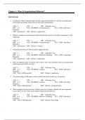
• Zone of ischemia: outermost ring of the MI; viable tissue if treated quickly; sign of past
MI; can cause T-wave inversion because of the hard time repolarizing
• Zone of Injury: middle ring of the MI; will always be affected but is not dead tissue; will
see ST elevation if transmural; sign of MI happening now
• Zone of infarction: dead and necrotic muscle; pathologic “Q-waves”
Transmural MI: “full thickness MI”; affects Endo-, Myo-, and Epicardium; will see Q-wave with
ST segment elevation
Subendocardial MI: multifocal areas; shows ST segment depression
, 12-lead ECG changes:
• Alterations in depolarization (systole)
o Change in QRS complex
• Alterations in repolarization (diastole)
o Change in ST segment (elevation or depression)
o Change in Q waves = Transmural MI
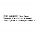
4 main arteries
• Left main coronary artery:
o “Widow maker”: blockage here is the patient who falls dead because of the
severity of blockage in this artery
o Feeds all of the left atrium and left ventricle so if you lose this artery you lose the
whole left side of your heart which is the working side
• Proximal Left Anterior Descending Artery:
o Anterior wall MI
o Can also call the “widow maker” it blockage is proximal enough because it
affects the left ventricle which is the most important
o ECG changes in leads V1, V2, V3, and V4
o Left ventricle pump failure →Cardiogenic shock → Death
o Will see hypoTN, tachycardia, tachypnea, decreased peripheral perfusion,
decreased pulses, skim mottling, decreased O2 sat, pink, frothy sputum (same as
left sided heart failure)
o Failure to pump forward backing up into lungs!!!!!!!
• Right coronary artery:
o Inferior wall MI
o ECG changes in leads II, III, aVf
o Common conduction problems
▪ RCA perfuses SA node in 50%; circumflex perfuses other 50%
▪ RCA perfuses AV node in 90%; circumflex perfuses other 10%
o Complications include:
▪ Bradycardia if SA node goes out
▪ Heart Block if AV node goes out
o Right ventricular infarction
▪ Proximal section of right coronary artery
▪ Can’t really pick up on 12-lead but can (not often) put electrodes on
backwards and put “R” on ECG
▪ Can cause cardiogenic shock because if right side isn’t pumping, the left
side doesn’t have anything to pump forward
▪ Will look like R side heart failure: JVD, edema, hepatomegaly, increase
CVP
• Circumflex artery
o Left lateral wall MI
o Only artery that doesn’t supply a ventricle
o Changes in I, aVL, V5, V6
Myocardial Infarction: “acute MI”; irreversible necrosis that results from an abrupt decrease or
total cessation of coronary blood flow to a specific area of the myocardium
• STEMI: “ST elevation MI”; usually caused by a clot and fibrinolytic therapy is used if not
contraindicated
• NSTEMI: “non-ST elevated MI; usually caused by plaque
• Manifestations:
o Tachycardia w/ or w/o ectopy: heart trying to compensate for decreased cardiac
output and ventricles are irritable because of hypoxia
o Bradycardia: if right sided MI
o Normotension or hypotension: hypoTN is left sided MI
o Tachypnea: from hypoxia, lungs are trying to get more O2
o Diminished heart sounds: decreased pressure causes valves to close softer which
causes decreased heart sounds
o S3: heart failure
o S4: hypertension
o Crackles: backup in lungs with left sided MI
o Pulmonary edema
o Air hunger
o Orthopnea
o Frothy sputum: HALLMARK SIGN
o Decreased CO
▪ Decreased peripheral pulses
▪ Slow capillary refill
o Decreased UO
o Decreased blood to brain
▪ Restlessness
▪ Confusion
▪ Agitation
▪ Anxiety
o Denial
o Anger
Patho:
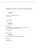
• Zone of ischemia: outermost ring of the MI; viable tissue if treated quickly; sign of past
MI; can cause T-wave inversion because of the hard time repolarizing
• Zone of Injury: middle ring of the MI; will always be affected but is not dead tissue; will
see ST elevation if transmural; sign of MI happening now
• Zone of infarction: dead and necrotic muscle; pathologic “Q-waves”
Transmural MI: “full thickness MI”; affects Endo-, Myo-, and Epicardium; will see Q-wave with
ST segment elevation
Subendocardial MI: multifocal areas; shows ST segment depression
, 12-lead ECG changes:
• Alterations in depolarization (systole)
o Change in QRS complex
• Alterations in repolarization (diastole)
o Change in ST segment (elevation or depression)
o Change in Q waves = Transmural MI
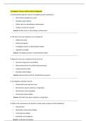
4 main arteries
• Left main coronary artery:
o “Widow maker”: blockage here is the patient who falls dead because of the
severity of blockage in this artery
o Feeds all of the left atrium and left ventricle so if you lose this artery you lose the
whole left side of your heart which is the working side
• Proximal Left Anterior Descending Artery:
o Anterior wall MI
o Can also call the “widow maker” it blockage is proximal enough because it
affects the left ventricle which is the most important
o ECG changes in leads V1, V2, V3, and V4
o Left ventricle pump failure →Cardiogenic shock → Death
o Will see hypoTN, tachycardia, tachypnea, decreased peripheral perfusion,
decreased pulses, skim mottling, decreased O2 sat, pink, frothy sputum (same as
left sided heart failure)
o Failure to pump forward backing up into lungs!!!!!!!
• Right coronary artery:
o Inferior wall MI
o ECG changes in leads II, III, aVf
o Common conduction problems
▪ RCA perfuses SA node in 50%; circumflex perfuses other 50%
▪ RCA perfuses AV node in 90%; circumflex perfuses other 10%
o Complications include:
▪ Bradycardia if SA node goes out
▪ Heart Block if AV node goes out
o Right ventricular infarction
▪ Proximal section of right coronary artery
▪ Can’t really pick up on 12-lead but can (not often) put electrodes on
backwards and put “R” on ECG
▪ Can cause cardiogenic shock because if right side isn’t pumping, the left
side doesn’t have anything to pump forward
▪ Will look like R side heart failure: JVD, edema, hepatomegaly, increase
CVP
• Circumflex artery
o Left lateral wall MI
o Only artery that doesn’t supply a ventricle
o Changes in I, aVL, V5, V6